Review

Pseudomonas Lipopeptide-Mediated Biocontrol: Chemotaxonomy and Biological Activity

1
Université de Reims Champagne Ardenne, Unité de Recherche RIBP EA4707 USC INRAE 1488, SFR Condorcet FR CNRS 3417, 51100 Reims, France
2
Department of Biological Sciences, Faculty of Science, Anchor University, Ayobo P.M.B 00001, Lagos State, Nigeria
3
Unit for Environmental Sciences and Management, Faculty of Natural and Agricultural Sciences, North-West University, Potchefstroom 2520, South Africa
4
Plant Pathology Unit, National Root Crops Research Institute (NRCRI), Umudike 440001, Abia State, Nigeria
5
Department of Pharmaceutical Biology, Institute of Pharmaceutical Sciences, University of Tubingen, Auf der Morgenstelle 8, 72076 Tübingen, Germany
6
Laboratory of Phytopathology, Department of Plants and Crops, Faculty of Bioscience Engineering, Ghent University, Coupure Links 653, B-9000 Ghent, Belgium
*
Author to whom correspondence should be addressed.
Molecules 2022, 27(2), 372; https://doi.org/10.3390/molecules27020372
Submission received: 1 December 2021 / Revised: 29 December 2021 / Accepted: 5 January 2022 / Published: 7 January 2022
(This article belongs to the Special Issue Biosynthesis and Biological Activities of Natural Products)

Abstract

:
Pseudomonas lipopeptides (Ps-LPs) play crucial roles in bacterial physiology, host–microbe interactions and plant disease control. Beneficial LP producers have mainly been isolated from the rhizosphere, phyllosphere and from bulk soils. Despite their wide geographic distribution and host range, emerging evidence suggests that LP-producing pseudomonads and their corresponding molecules display tight specificity and follow a phylogenetic distribution. About a decade ago, biocontrol LPs were mainly reported from the P. fluorescens group, but this has drastically advanced due to increased LP diversity research. On the one hand, the presence of a close-knit relationship between Pseudomonas taxonomy and the molecule produced may provide a startup toolbox for the delineation of unknown LPs into existing (or novel) LP groups. Furthermore, a taxonomy–molecule match may facilitate decisions regarding antimicrobial activity profiling and subsequent agricultural relevance of such LPs. In this review, we highlight and discuss the production of beneficial Ps-LPs by strains situated within unique taxonomic groups and the lineage-specificity and coevolution of this relationship. We also chronicle the antimicrobial activity demonstrated by these biomolecules in limited plant systems compared with multiple in vitro assays. Our review further stresses the need to systematically elucidate the roles of diverse Ps-LP groups in direct plant–pathogen interactions and in the enhancement of plant innate immunity.

Graphical Abstract

1. Introduction

The Pseudomonas genus is ubiquitous and comprises species which are well known phytopathogens, such as P. syringae, or opportunistic human pathogens, such as P. aeruginosa, but also host members associated with water, soil and plant surfaces [1]. Pseudomonas spp. are well adapted to growing in the rhizosphere and are well suited for biocontrol and growth promotion [2]. Thus, the use of fluorescent Pseudomonas spp. as potential biopesticides has gained attention over the last decade. These bacteria are of particular interest because of their enormous metabolic versatility and wide adaptation across environmental gradients [3].
Based on phylogenomic and Multi Locus Sequence Analyses (MLSA), the Pseudomonas genus has been delineated into 453 species (https://lpsn.dsmz.de/genus/pseudomonas; accessed on 18 December 2021) which are distributed across three lineages (P. fluorescens, P. aeruginosa and P. pertucinogena), several groups (G) and subgroups (SG) [4,5,6,7,8]. Most biocontrol strains have been described within the P. fluorescens group comprising among others, the P. fluorescens SG, P. koreensis SG, P. chlororaphis SG, P. jessenii SG, P. mandelii SG and P. corrugata SG. Additionally, several biocontrol strains are positioned within the P. putida and P. syringae groups. These disease-suppressing pseudomonads were isolated from several sources ranging from the healthy plant rhizosphere [9,10,11], plant rhizosphere [12,13,14,15], phyllosphere [16,17], bulk soil [15] and suppressive soils [10,18]. The commonality among well-studied biocontrol strains is their capacity for secondary metabolite production including siderophores, lipopeptides (LPs), hydrogen cyanide, bacteriocins and certain antibiotics such as phenazines, 2,4-diacetylphloroglucinol (DAPG), pyrrolnitrin and pyoluteorin [3,19].
Examples of commercially available Pseudomonas-based bioprotectants include fungicides such as Cedomon and Cerall (P. chlororaphis MA342) both targeting seed-borne pathogens of cereals, Spot-Less (P. aureofaciens strain Tx-1) for management of fungal diseases on lawns and grasses, and Howler (P. chlororaphis AFS009) useful in the management of Rhizoctonia, Pythium, Fusarium, Phytophthora, Colletotrichum spp. in fruits, vegetables and ornamentals [19]. A detailed list of commercial bioprotectants based on Pseudomonas in Europe and USA, including their usage, and target crops/applications/pathogens have been enumerated in a recent review [19].
Lipopeptides are bacterial metabolites consisting of a peptide part attached to a fatty acid tail [1]. Most beneficial LPs are cyclized although linear LPs have also been described [20,21]. LPs have drawn remarkable interest because of their broad-spectrum antimicrobial and ecological functions. These multiple functions include biofilm formation and colonization of surfaces, quorum sensing, cell motility, soil remediation, anti-oomycete, antiviral, antifungal, antibacterial, herbicidal, insecticidal, antiprotozoal and anticancer properties [3,22,23,24,25,26,27].
In the past decade, the role of secondary metabolites, especially of LPs contributing to the biocontrol capability of Pseudomonas spp., has been increasingly studied [1,28]. This research wave was triggered by the increasing novelty of LPs that were being structurally and functionally characterized. On the one hand, several new LPs have been characterized within Pseudomonas groups [6,11,29] while the functions of some LPs have been recently characterized using insertion or deletion mutagenesis, crude LP extracts or purified LPs. Besides the review that highlighted the advances in Pseudomonas biocontrol [20], three recent reviews summarized so far the role of biosurfactants (mainly Bacillus LPs and rhamnolipids) in plant disease protection [30], described diverse elicitors of plant immunity produced by beneficial bacteria [31] and the use of Pseudomonas spp. as bacterial biocontrol agents to control plant disease [19]. The current review provides a summary of LP-producing biocontrol strains situated within specific Pseudomonas groups and highlights the taxonomy–molecule specificity of biocontrol Ps-LPs. Moreover, future areas of research and methodologies are proposed in order to accelerate our understanding of Ps-LP-based biocontrol and Ps-LP-pathogen interactions. Other aspects of LP-mediated plant–pathogen interactions are also discussed.

2. Methodology

In this review, bibliometric data were extracted from the SCOPUS database (https://www.scopus.com/; accessed on 20 February 2021) using the following specific keywords viscosin OR amphisin OR bananamide OR cocoyamide OR orfamide OR tolaasin OR syringomycin OR syringopeptin OR xantholysin OR putisolvin OR entolysin OR “cyclic lipopeptide” OR “cyclic lipopeptides” OR “CLPs” OR “lipopeptide” OR “lipopeptides” OR “LPs” AND Pseudomonas from which 118 documents were obtained. The bibliometric analysis was constructed using the VOSviewer processing software (v1.6.9., Leiden University, Leiden, The Netherlands).
A Comparative Genomic Blast Atlas was created using Pseudomonas genomes of 35 lipopeptide-producing strains, extracted from the National Centre of Biotechnology Information (NCBI) website. P. fluorescens Pf0-1 was chosen as the reference genome while representative LP-producing strains, selected from diverse Pseudomonas taxonomic groups/subgroups were uploaded as related genomes (Appendix A). As results, regions will be displayed where there is a similarity between the reference genome and one of the related genomes. Multi-Locus Sequence Analysis (MLSA) phylogenetic analysis was also conducted using the MEGAX software.
The literature search was conducted by accessing several databases including Scopus, PubMed, Web of Science, SpringerLink, Google Scholar and ResearchGate. A total of 126 articles and three book chapters were used. Schematic illustrations were drawn using the Biorender software.
For clarity, a series of detailed steps employed in writing this review is represented as a flowchart [32] (Figure 1).

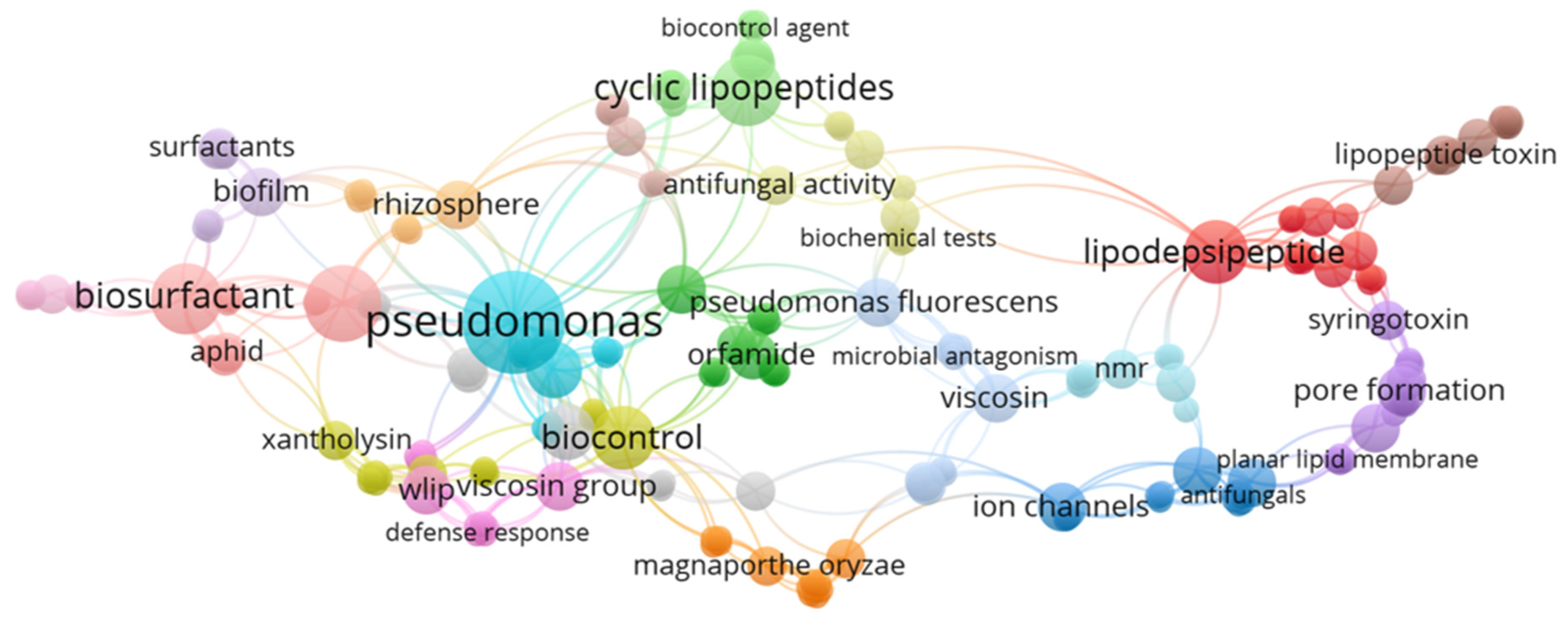
3. Network Analysis Showing the Distribution of Pseudomonas LP-Related Articles

The network analysis showed the distribution of articles related to cyclic lipopeptides, which helped to highlight the relationship between the keywords found and allowed a comprehensive perspective of the current research in this area (Figure 2). Clearly, these research areas will be enumerated in detail within this review.

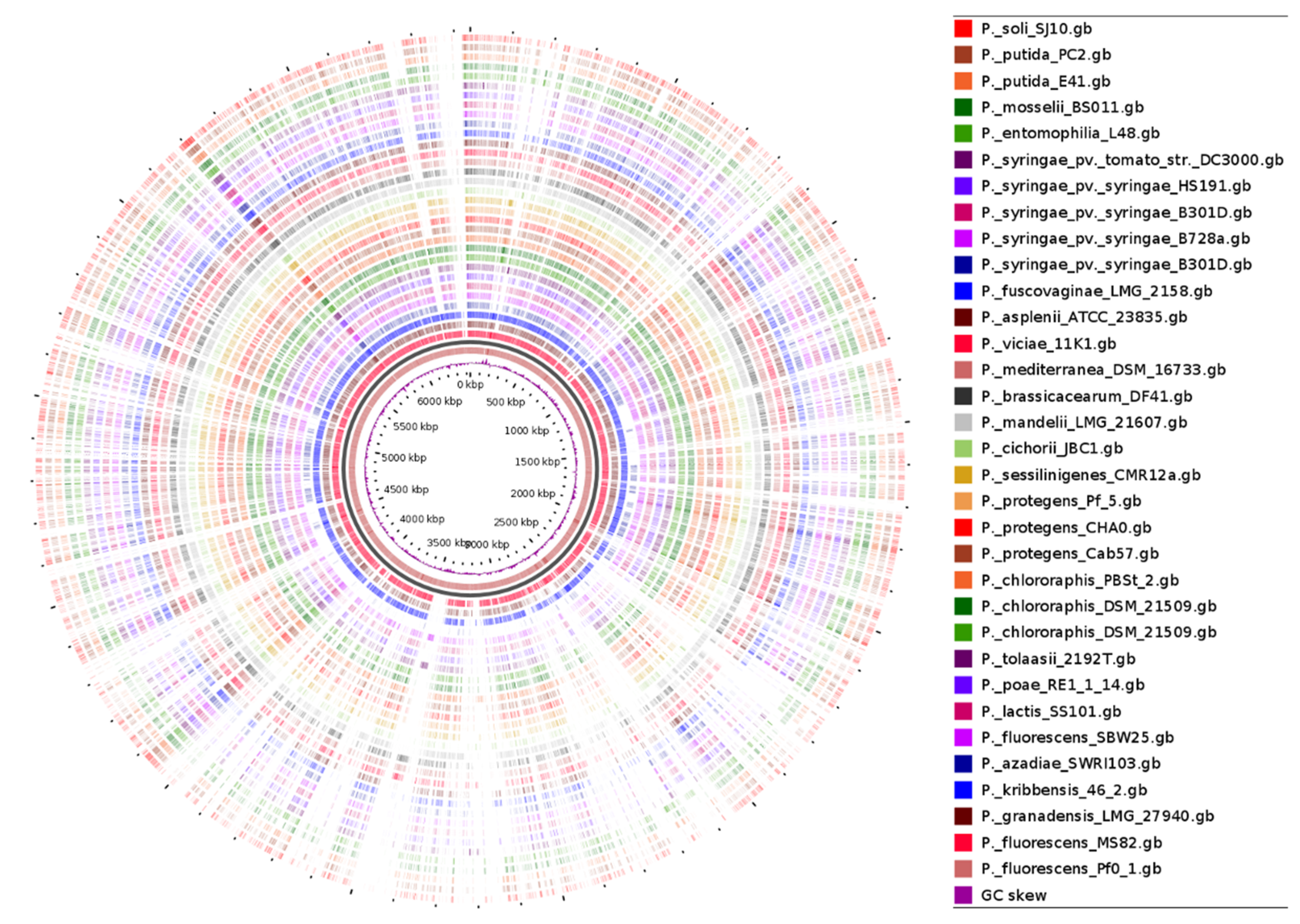
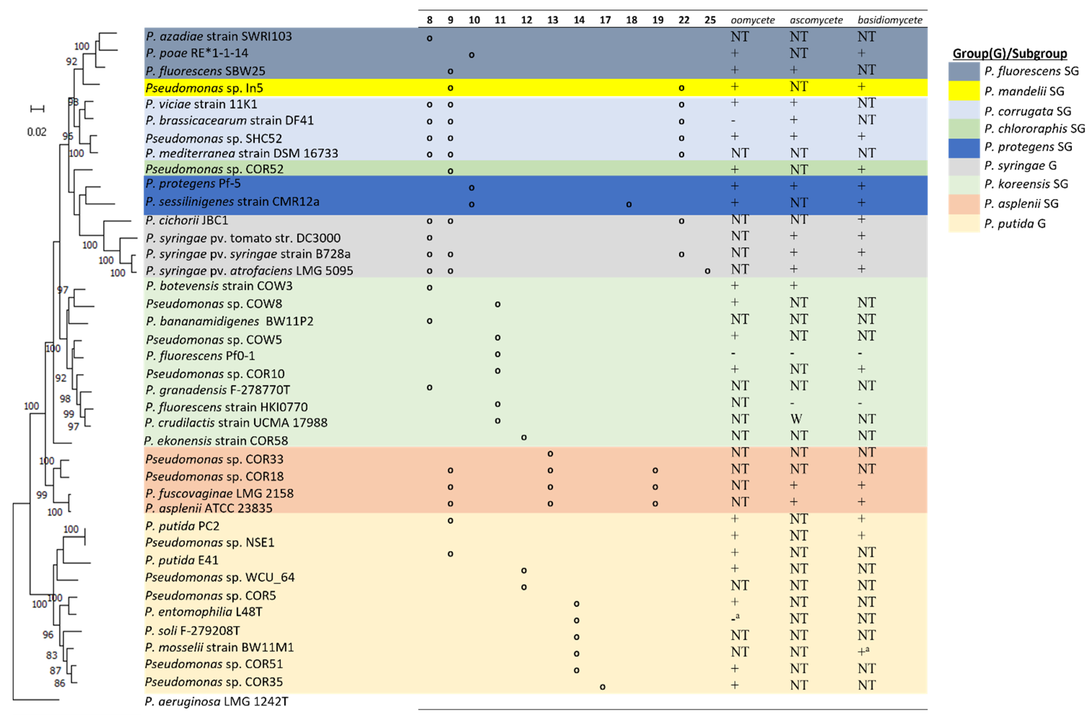
4. Genome Comparison of Selected Lipopeptide-Producing Pseudomonas spp.

A previous study provided the phylogenomic analysis of the Pseudomonas genus based on the genomes of the type strains of 163 described species and compared these type strain genomes to those of 1223 Pseudomonas genomes in public databases [7]. Results showed that 400 of those 1223 genomes were distinct from any other type strain suggesting that the Pseudomonas genomic diversity had been grossly underrepresented by the type strains. Furthermore, a detailed comparative genome analysis of ten strains within the Pseudomonas fluorescens group highlighted the enormous diverseness of this group and the capacity of the variable genome to adapt individual strains to their distinct lifestyles and functional capacities [3]. Here, using the P. fluorescens Pf0-1 as a reference genome, we compared the genome of 32 lipopeptide-producing Pseudomonas strains affiliated with the P. koreensis, P. fluorescens, P. mandelii, P. corrugata, P. asplenii, P. chlororaphis, P. protegens, subgroups including the P. putida and P. syringae groups. By comparing the protein coding sequences (CDS) of reference to query genomes, a Blast Atlas was generated which showed the close relatedness of other members of the P. koreensis group (P. fluorescens MS80, P. granadensis LMG 27,940 and P. kribbensis 46-2) to the reference genome P. fluorescens Pf0-1 (Figure 3). Clearly, these genomes are highly variable and distinct. Detailed comparative gene identities are presented in Table S1.

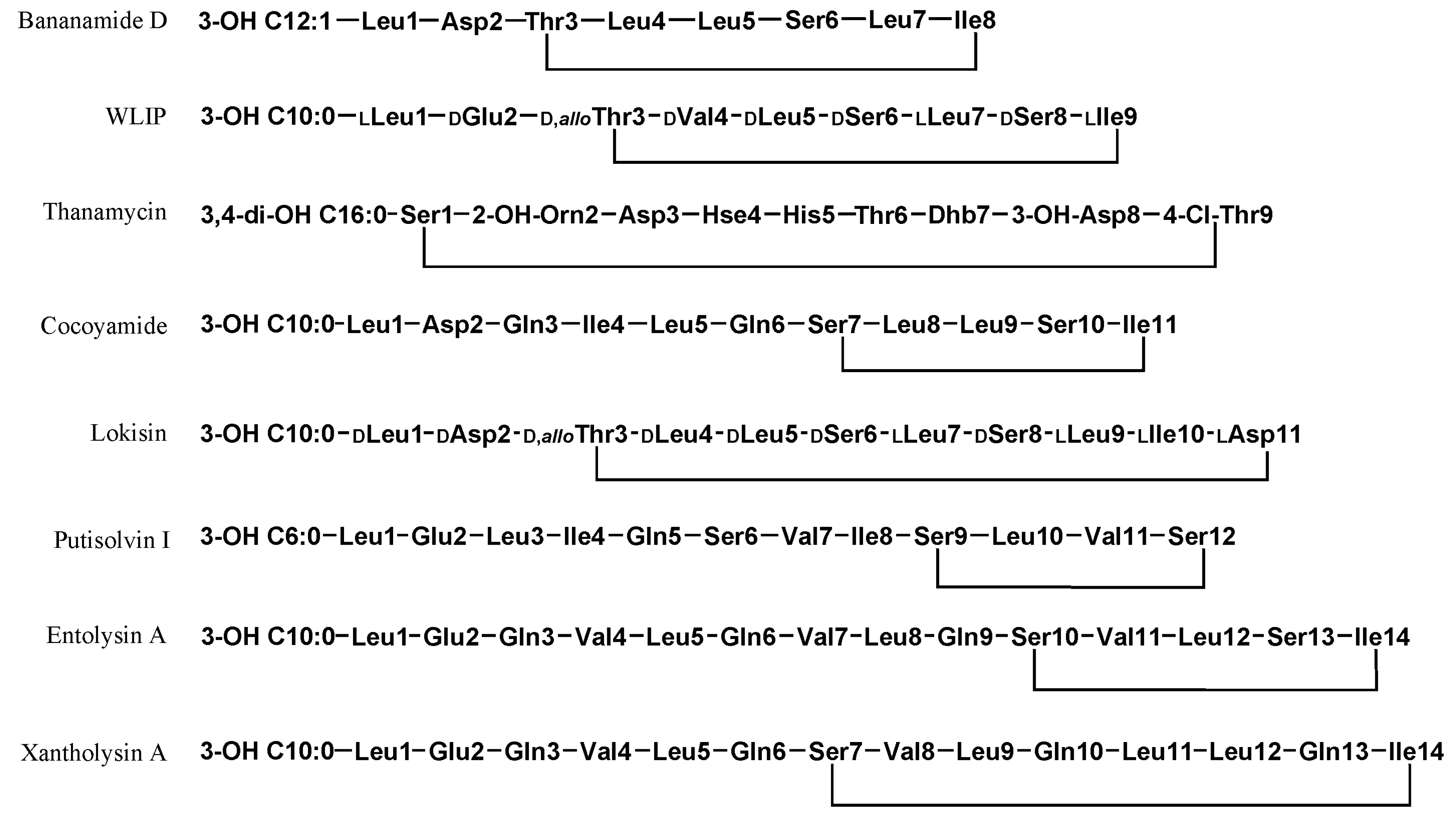
5. Chemical Diversity of Beneficial Pseudomonas LPs

Most beneficial LPs have been predominantly characterized from strains affiliated with the P. fluorescens and P. putida group. The chemical diversity of Pseudomonas LPs has been detailed in two recent reviews [1,21]. Table 1 shows the diversity of beneficial LPs and presents the discovery of similar LPs from diverse strains, countries, niches and environments. Not all LPs listed have been functionally characterized, however, the disease suppressive capacity of their producing strain(s) has been established on specific plant hosts thus indicating non-virulence. Clearly, the P. koreensis subgroup presents the highest diversity of LP families and individual members, including variants. This SG is characterized by at least six amphisin group members alongside the novel rhizoamide, the bananamide group comprising six variants and the cocoyamide/gacamide group. Moderate LP diversity is showcased by the P. fluorescens SG while the P. protegens SG comprises various orfamide variants A-H and the poaeamide LPs. Lastly, the P. putida group contains four described LP types: entolysin, putisolvin, xantholysin, WLIP and a novel 17AA LP named N8. Figure 4 shows the chemical structures of representative biocontrol LPs that have been characterized.

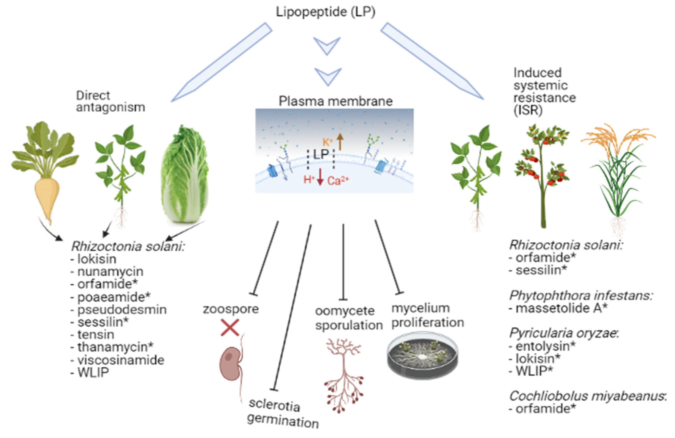
6. Pseudomonas LPs: Broad Spectrum Arsenals for Biological Control of Plant Pathogens

The Pseudomonas genus being a tremendous source of diversity, uniquely houses new strains and species [35]. The biocontrol activity displayed by these strains, corresponding LP mutants or crude as well as purified LPs has been summarized in Table 2. In this section, these active LPs will be discussed according to the taxonomic affiliation of producing strains and their respective LP groups.

6.1. The P. fluorescens SG: Houses the Viscosin Group, Certain Members of the Tolaasin Group and the Poaeamide Producer

Until recently, the P. fluorescens SG appeared to have strains producing the highest LP diversity. Besides the tolaasin group, LPs produced within this Pseudomonas subgroup mainly belong to the viscosin group. Members of this group comprise an oligopeptide of 9AA, differing from each other mainly by the presence of a Leu, Ile or Val at positions 4 and 9, and the 3-hydroxy-fatty acid tail length (10 to 12 carbons) [21]. Members of this group include viscosin (vis), viscosinamide (vsm), massetolide (mass), pseudodesmin (pdm), pseudophomin (psm) and the white line inducing principle (WLIP). Viscosin has been described from multiple sources; strains causing head rot of broccoli [84], marine environment [85], and the sugar beet plant phyllosphere [35,86]. Massetolide A has been isolated from strains associated with the leafy red sea algae surface [85] and from the wheat rhizosphere isolate, P. lactis SS101 [34]. Within the P. fluorescens SG, the WLIP was first reported in P.reactans” [69] and subsequently detected in several Pseudomonas strains including P.reactans” NCPPB1311 [42] P. putida RW10S2 (now called P. promisalinigenes RW10S2), a rice rhizosphere isolate [67], the biocontrol strain P. chlororaphis PB-St2, an isolate from sugarcane stems [69] and from the cocoyam rhizosphere isolate, NSE1 [11,68]. Two viscosinamide producers, P. fluorescens DR54 and Pseudomonas sp. A2W9.4, were isolated from sugar beet [37,87] and cocoyam rhizosphere [38], respectively. Pseudophomins A and B are produced by strain BRG100, a green foxtail rhizosphere isolate [39]. The most recently reported member of this group, pseudodesmins A and B, were isolated from Pseudomonas bacteria obtained from the mucus layer in the skin of the black belly salamander [88] and also recently characterized from the cocoyam rhizosphere in Cameroon [38].
Until recently, LPs belonging to the viscosin family have been most researched for biocontrol capabilities against plant pathogens including bacteria, fungi and oomycetes. In vitro assays using cell cultures, cell-free culture supernatants of P. fluorescens SBW25 or purified viscosin showed an efficacy in the immobilization and subsequent lysis of zoospores of Phytophthora infestans [35]. The biocontrol potency of SBW25 due to viscosin against P. infestans was further strengthened in tests involving viscosin-deficient mutants. Similar positive effects of local and induced systemic resistance in the control of P. infestans-mediated late blight of tomato was obtained using cell cultures and cell-free supernatants of the massetolide producer (SS101), Tn5 massetolide mutants and purified massetolide A [72]. In contrast, the massetolide production by P. lactis SS101 was not required to suppress the complex Pythium populations on apple and wheat [89]. Furthermore, a salicylic acid-dependent resistance response was successfully induced by SS101 on Arabidopsis thaliana against Pseudomonas syringae pv. syringae [73]. However, the probable role of massetolide in the observed induced systemic resistance (ISR) was not investigated.
Although no LP mutants have been constructed in pseudophomin, pseudodesmin and viscosinamide-producing strains, the bioactivity of these LPs has been demonstrated against Gram-positive/negative bacteria and/or plant pathogens. Pseudophomins A and B showed antifungal activity against Leptosphaeria maculans and Sclerotinia sclerotiorum [39] and were also antagonistic towards several human pathogenic Gram-positive bacteria [88].
In vitro tests with viscosinamide (Vsm) against P. ultimum and R. solani showed a reduction in biomass and radial growth of mycelium [14,36,70]. In situ application of Vsm resulted in decreased oospore formation and sclerotia formation in P. ultimum and R. solani, respectively [36,37,70,71,87]. In a more recent study, in vitro tests with varying Vsm concentrations showed that nanomolar levels caused hyphal distortion and branching of P. myriotylum including hyphal lysis at 1 µM [38]. Similarly, in in vitro tests against R. solani AG2-2, nanomolar to micromolar levels of Vsm resulted in blockage of hyphal formation, hyphal distortion and pronounced LP evasion phenotypes [38].
WLIP, a LP produced by strains belonging to the P. fluorescens SG, P. chlororaphis SG and the P. putida G, was found to inhibit the brown blotch disease on Agaricus bisporus caused by P. tolaasii [90]. In a related study, cell-free crude extract containing WLIP from P.reactans” SPC 8907 inhibited the same pathogen [91]. Furthermore, WLIP production by P.reactans” NCPPB1311 demonstrated antagonism against Erwinia carotovora subsp. carotovora (now called Pectobacterium carotovorum) and Agaricus bisporus [42]. Subsequent biological screening against fungi and bacteria indicated that WLIP is more effective against Gram-positive bacteria than Gram-negative [42]. In a separate study, tests with purified WLIP (from P. chlororaphis Pb-St2) did not show efficacy in antifungal tests involving R. solani AG2-2 and AG4 isolates [69]. In contrast, WLIP obtained from Pseudomonas sp. NSE1 showed excellent efficacy when tested against the same AG2-2 pathogen but using a different experimental set up [38]. Thus, in comparative studies, the need for a consistent approach when comparing compound efficacies cannot be overemphasized. Like results obtained with R. solani AG2-2, variable WLIP concentrations resulted in complete hyphal destruction of P. myriotylum [38] Moreover, Tn5 insertion WLIP mutants of P. promysalinigenes RW10S2 led to an antagonism against plant pathogenic Xanthomonas species and inhibited the growth of several Gram-positive bacteria in vitro [67].
Purified poaeamide solution caused immobilization and subsequent lysis of Phytophthora capsici and P. infestans zoospores within 1 min exposure at CMC concentrations of 50 μg/mL [41]. In contrast, cell-free culture supernatants of the WT strain did not cause these responses in the pathogens tested. When grown in direct contact with the 50 μg/mL LP solution, the inhibitory effect on dry weight mycelial biomass was observed in P. capsici, R. solani and P. infestans whereas a similar effect was only accomplished in P. ultimum at 250 μg/mL. Furthermore, an inhibitory effect was observed in the mycelial fresh weight for the same pathogens except for P. infestans.
The composition and length of the peptide chain of the tolaasin group ranges between 19 to 25 amino acids with the lipid tail comprising of 3-HDA or 3-hydroxyoctanoic acid (3-HOA). Purified tolaasin I from P. tolaasii NCPPB2192 inhibited the growth of fungi namely Agaricus bisporus, Lentinus edodes, Pleurotus spp. and Gram-negative bacteria belonging to the genera Erwinia, Agrobacterium, Xanthomonas, Escherichia and Pseudomonas [42]. Sessilins also belong to the tolaasin group of LPs but are produced by P. sessiligenes CMR12a, an affiliate of the P. protegens SG.

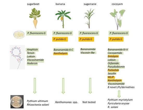
6.2. The P. koreensis SG: LP Cocktail Comprising Amphisin, Bananamide and Cocoyamide Groups

The P. koreensis group is a LP cocktail group comprising at least three different LP groups. In recent years, several novel LPs and derivatives have been characterized from strains situated within this subgroup. The amphisin LP group are produced within this species and comprise members including amphisin, anikasin, arthrofactin, lokisin, milkisin and tensin (Table 1). Tensin was derived from the P. fluorescens strain 96.578. Amphisin-, lokisin- and tensin-producing Pseudomonas strains demonstrated a high level of antagonism against P. ultimum and R. solani [51]. Subsequent in vitro assays using these three compounds confirmed their antagonism against these two pathogens [14,51]. Recent in vitro studies using a lokisin deletion mutant of the P. koreensis S150 strain showed reduced activity against Phytophthora nicotianae and a complete loss of inhibition against R. solani [49]. Additionally, purified lokisin lysed the mycelia of the cocoyam root rot pathogen, P. myriotylum [10]. Pseudomonas sp. DSS73 inhibited the root pathogenic fungus R. solani partially because of amphisin production [43]. A latest LP addition is rhizoamide A (formerly described as N2) which has been shown to cause hyphal lysis of P. myriotylum at low concentrations [10,11]. Full structural and functional analysis of rhizoamides A and derivatives B-D will be described elsewhere.
The first members of the bananamide group, bananamides I-III, were described from the banana rhizosphere isolate BW11P2 [52]. More so, a closely related LP (yet to be chemically characterized) is reportedly produced by a wheat rhizosphere isolate, P. azadiae SWRI103 [92]. So far, the antimicrobial and biocontrol activities of bananamides I-III have not been demonstrated. Another member of this group, bananamide D, was described from cocoyam rhizosphere isolates, COW3 and COW65 [29]. Like the first producer, COW3 produced other variants namely bananamides E, F and G. Strain COW3 suppressed cocoyam root rot disease (CRRD) in soil assays [10] while in in vitro tests, bananamides D-G inhibited the growth of P. myriotylum in a dose-dependent manner and further induced hyphal branching and leakage [29]. In the same study, the growth of P. oryzae was significantly inhibited by bananamide A, while other derivatives only displayed a mild effect. Lastly, mycophagous behaviour of bananamide D producers was observed against P. oryzae although it is unclear whether this is directly or indirectly due to LP production.
Cocoyamide A/Gacamide A, belonging to a new LP group, was first described from the cocoyam rhizosphere isolate Pseudomonas sp. COW5 [10] and from the GacA complemented P. fluorescens Pf0-1 strain [53]. Pseudomonas sp. COW5 effectively protected cocoyams from the CRRD while 10 µM of purified cocoyamide A was sufficient to cause lysis of the host-adapted cocoyam pathogen, P. myriotylum [10]. Purified gacamide was also described as having a moderate, narrow-spectrum antibiotic activity against clinical bacterial isolates [53].

6.3. The P. protegens SG: Home to Multiple Orfamide Derivatives and Sessilins

Orfamides were first extracted from P. protegens Pf-5 and subsequently from P. protegens CHA0, P. protegens F6 [93], P. sessiligenes CMR12a [59], Pseudomonas sp. Cab57 [94], P. aestus CMR5c, P. fluorescens Wayne1R, Pseudomonas spp. CMAA1215 and PH1b [56]. Multiple orfamide derivatives are produced by different strains including orfamides A-G (Table 1); strains Pf-5 and CHA0 both produce orfamides A, B and C whereas CHA0 produces an additional G derivative. More so, both CMR12a and CMR5c produce orfamides B, D, and E while the latter also produced F and G derivatives. Using the water agar–LP droplet assay, 100 µM of orfamides (A, B and G) caused hyphal branching indicative of mycelium growth inhibition in R. solani AG-4 HGI [56,75], contrasting negative results were obtained for the same pathogen when the agar diffusion assay method was used [79]. Orfamide B increased hyphal branching of R. solani AG2-1, the pathogen causing damping-off of Chinese cabbage [75]. At concentrations of 25 µM or higher, orfamides A, B and G caused zoospore lysis of P. ultimum and Phytophthora porri CBS 127099 [56]. Previous studies also showed that orfamide A and viscosin family of LPs, WLIP and viscosinamide can lyse zoospores of the oomycete pathogen, Phytophthora ramorum [35,71,79]. Soil assays with orfamide biosynthesis mutants revealed that orfamide B, produced by P. sessiligenes. CMR12a, work synergistically with phenazines and sessilins to suppress R. solani AG4-mediated root rot of bean, damping-off of Chinese cabbage caused by R. solani AG2-1 [75] and Pythium root rot of cocoyams [77]. Besides its efficacy against plant pathogens, orfamide A showed dose-dependent insecticidal mortality against aphids [93] and was reported to be a major determinant in the oral toxicity of Pf-5 against Drosophila melanogaster [95]. Additionally, experiments conducted using mutants and purified orfamide A showed that orfamide A could not elicit induced systemic resistance against the rice blast pathogen, P. oryzae but successfully elicited ISR against Cochliobolus miyabeanus, the causal pathogen of brown spot disease. Interestingly, introducing high inoculum of strain CHA0 successfully mediated ISR against C. miyabeanus on rice while this could not be achieved with strain CMR12a [78].
Sessilin, produced by the cocoyam rhizosphere isolate P. sessiligenes CMR12a, is structurally related to tolaasin and only differs from tolaasin I by one amino acid. Direct application of crude sessilin extracts resulted in vacuole formation and subsequent lysis of the mycelia of P. myriotylum [77]. Besides sessilin, CMR12a produces orfamide and two phenazine derivatives, PCA and phenazine-1-carboxamide (PCN). Using mutant analysis and soil assay experiments, sessilin was shown to be involved in the suppression of bean root rot due to R. solani AG2-2 [74], AG4 [75] and in the control of damping-off disease of cabbage caused by R. solani AG2-1 [75].

6.4. P. chlororaphis SG: Pseudodesmin, WLIP and Uncharacterized Viscosin Group LPs

The P. chlororaphis group comprises strains from the soil and the rhizosphere of diverse plant hosts [19]. Multiple secondary metabolites have been reported in this group [96] including the production of viscosin group lipopeptides. The sugarcane stem isolate, P. chlororaphis subsp. aurantiaca, produces WLIP but the compound did not display antifungal or antibacterial activity [69]. Viscosin group LPs have been described although their full structure and bioactivity potential is yet to be deciphered [96]. Furthermore pseudodesmin has been characterized from the P. chlororaphis-grouped cocoyam rhizosphere isolate, Pseudomonas sp. COR52 [38], thus its antimicrobial activity will be discussed here. Using a broth microdilution method, the antimicrobial activity of synthetic pseudodesmin (Pdm) was shown against six Gram-positive bacterial pathogens [97]. In this study, no antifungal effect was recorded against Candida albicans and Aspergillus fumigatus, suggesting that this compound is mainly active against Gram-positive bacteria. However, a follow up study showed the antifungal activity of this LP against the tropical cocoyam root rot pathogen, P. myriotylum, and the bean root rot pathogen, R. solani AG2-2 [38]. 100 nM and micromolar concentrations of Pdm inhibited the mycelial growth of P. myriotylum, effected hyphal distortions and branching, while specific Pdm concentrations (100 nM and 25 µM) resulted in a unique hyperbranching phenotype [38]. When similar concentrations were tested against R. solani AG2-2, multiple hyphal changes were also observed including growth inhibition, hyphal distortion, blockage and lysis.

6.5. P. mandelii SG, P. asplenii SG and P. corrugata SG: Thin Borderline between Pathogenic and Beneficial LPs I

In contrast to the P. mandelii SG, strains affiliated with the P. asplenii and P. corrugata SGs are predominantly plant pathogenic species which may also double up as plant beneficial bacteria [28]. The production of multiple LPs is a key characteristic of strains in these groups and in most cases, the complementary and synergistic roles played by the LPs in phytotoxicity, virulence or biocontrol have been reported and discussed. The antimicrobial activities of associated purified as well as crude LPs produced within these taxonomic groups have been recently summarized [28]. Here, we succinctly highlight the roles of beneficial LPs in these otherwise plant-pathogenic groups. Furthermore, we present a comparison of Pseudomonas strains taxonomy, the AA composition of their LPs and reported activity against three plant pathogenic classes (Figure 3).
P. mandelii: Nunamycin and nunapeptin, LPs produced by P. fluorescens In5 were reported to be key components for the biocontrol activity of In5 [18]. Differential inhibition was exhibited by both LPs against similar plant pathogens; nunamycin inhibited mycelial growth of R. solani AG3 but was not effective against Pythium aphanidermatum whereas, with nunapeptin, the opposite scenario played out—the later pathogen was inhibited in vitro or suppressed in soil assays with tomato whereas, R. solani AG3 was not [18].
P. corrugata: Thanamycin, brabantamide A and thanapeptin LPs are produced by Pseudomonas sp. SH-C52, a R. solani-suppressive soil isolate [60,81,98]. Experiments using mutants constructed in the thanamycin BGC and with the pure compound revealed the involvement of thanamycin in the biocontrol of SH-C52 against Sclerotium rolfsii on groundnut, R. solani on sugarbeet and the Gram-positive bacterium B. megaterium, but little activity against oomycete pathogens and certain Gram-negative bacteria. Brabantamide showed activity against Gram-positive bacteria such as Staphylococcus aureus and Arthrobacter crystallopoietes [99,100] and up to 50 µM was necessary to exhibit anti-oomycete activity against P. capsici and P. infestans [60]. However, phospholipases of the late blight pathogen, P. infestans were affected upon overnight co-incubation with 5 µM of brabantamide. Comparison of two Tn5 thanapeptin BGC mutants with the WT strain SH-C52 showed that thanapeptin is active against oomycetes but is not antifungal [60]. In vitro tests using purified compounds of seven thanapeptin derivatives revealed substantial differences in anti-oomycete activity such that compounds with the lowest mass had the strongest activity. For closely related LPs, syringopeptin (produced by P. syringae G isolates) and corpeptin (produced by P. corrugata SG strains), which are closely related LPs, no anti-oomycete activity has been reported [101,102].
Similar to reported LP producers in the P. corrugata SG, brasmycin and braspeptin LPs are produced by Pseudomonas sp. 11K1 [62]. A brasmycin deletion mutant lost inhibition activity against Botryosphaeria dothidea whereas the braspeptin mutant exhibited reduced antifungal activity. Both CLPs were not antibacterial against Xanthomonas oryzae RS105. Co-inoculation of purified sclerosin, produced by P. brassicacearum DF41 with Sclerotinia sclerotiorum, showed inhibition of ascospore and sclerotia germination but was not active against zoospores of P. infestans [61].
P. asplenii: The P. asplenii subgroup consists of P. fuscovaginae (Pfv) and P. asplenii species comprising rice-infecting pathogens causing sheath rot and grain discoloration symptoms [103]. Disease surveys of rice-infected fields in tropical ecologies (Philippines) have revealed several Pfv-related strains which form a distinct population which differ from the type Pfv strains and were thus referred to as being Pfv-like [103]. Typically, Pfv and Pfv-like species produce syringotoxins, fuscopeptin A (FP-A) and fuscopeptin B (FP-B) which are key virulence factors in rice sheath rot infection [103,104,105,106]. Although Pfv and related strains are not typically biocontrol strains, the antimicrobial activity of its associated LPs have been reported. Like syringotoxin, the purified fuscopeptin inhibited the growth of B. cinerea and R. pilimanae [104].
Pfv-like strains Pseudomonas spp. COR33 and COR18 were isolated from the tropical cocoyam rhizosphere in Cameroon [10]. Similar to known Pfv-like strains, COR18 produces multiple LPs; a novel cyclic LP named N5, having 13AA with eight in the macrocycle (13:8) and at least a novel peptin-like LP named N7 [11]. Similarly, COR33 produces N5 but differs from typical Pfv-like strains since it does not produce multiple LPs. In soil assays, strain COR33 suppressed the cocoyam root rot disease caused by P. myriotylum. The structural and functional characterization of COR18 and COR33 LPs vis-à-vis those of Pfv LPs will be published shortly. In a recent review paper, LP-13 (identical to N5) was reported to be produced by Pfv strains in addition to fuscopeptin, syringotoxin and cryptic BGCs [28]. The clustering of COR18 (a multiple LP producer) with Pfv-like strains and the distinct separation of COR33 (a single LP producer) from this group provided the possibility to investigate LP evolution and function within the P. asplenii group. Although biocontrol assays on cocoyams or other crops are yet to be conducted using COR18, it is unlikely to be phytotoxic on cocoyam since it was isolated from the healthy cocoyam rhizosphere.

6.6. P. syringae G: Thin Borderline between Pathogenic and Beneficial LPs II

Popular biocontrol P. syringae group isolates include the P. syringae pv. syringae B359 (alternatively named B497 or HS191), P. syringae pv. syringae B301 and P. syringae ESC-10 and ESC-11. Strains ESC-10 and ESC-11 effectively protected both lemons and oranges against green and blue molds caused by Penicillium digitatum and Penicillium italicum, respectively. The superior efficacy of P. syringae strain ESC-10 in controlling postharvest pathogens on citrus crops led to the development of Bio-Save, an EPA registered product containing ESC-10 as its active ingredient [83]. The application of Bio-Save reduced green and blue mold incidence on lemons and oranges by 87.9% and 58.6%, respectively. ESC-11 is used for the control of postharvest pathogens on apple and pear. Both ESC-10 and ESC-11 are commercialized as Bio-SAVE 10 and Bio-SAVE 11, respectively [83]. However, the biocontrol capacity of these strains is not due to LPs.
Syringomycin, syringotoxin and syringopeptin are phytotoxins produced by P. syringae which generally induce necrosis [107]. These LPs are known to act as virulence factors and facilitate disease severity when produced. The peptide portion of syringopeptin contains either 22 (SP22) or 25 (SP25) amino acids that are mainly hydrophobic. SP25 is produced by strains isolated from infected millet (B359) and citrus (B427), while SP22 is produced by a P. syringae isolate from pear (B301) and variants by strains obtained from different hosts. Besides enhancing virulence of the producing pathogens, purified syringomycin E (SR-E), syringotoxin (ST) and two forms of syringopeptin (SP22-A and SP25-A) exhibited antimicrobial activity against specific Gram-positive bacteria and fungi. Although the growth of Bacillus megaterium was inhibited by 1.56 µM SP22-A and 3.12 µM SP25-A, SR-E and ST had no effect [82]. With respect to fungi, the yeast Rhodotorula pilimanae was most sensitive to SR-E but was also inhibited by SP22-A and SP25-A. The plant pathogenic ascomycete B. cinerea was inhibited by 1.6 µM SP25-A, 12.5 µM SP22-A, 18.7 µM SR-E and 25 µM ST [82]. Despite having similar spore-forming characteristics in artificial membrane bilayer assays, these different P. syringae toxins have distinct antimicrobial activities.

6.7. P. putida G: Beneficial LPs with Broad-Spectrum Targets

Within the P. fluorescens lineage, the P. putida group is the second largest, containing about 69 species that occupy diverse ecological niches [6,7,27]. Besides their role in plant growth promotion and soil remediation, P. putida strains have functions in direct antagonism and ISR against plant pathogens [108,109]. So far, four main lipopeptides belonging to diverse families, have been described from strains within this group [20,110] (Table 1). Among the P. putida LPs is the WLIP whose bioactivity has been described in this review as a member of the viscosin group, within the P. fluorescens SG.
Following the discovery of the WLIP, putisolvins I and II have only been described from P. putida PCL 1445, isolated from a site polluted with polycyclic aromatic hydrocarbons [111] and P. putida 267, isolated from the black pepper rhizosphere [65]. More recently, putisolvins III, IV and V were described from cocoyam rhizosphere isolates Pseudomonas spp. COR19, COR55, NNC7, WCU_60, WCU_64 [10,11] and in P. putida LMG 11722T, the type strain of P. fulva [110]. Additionally, the prominent plant -growth promoting and -disease suppressing strain, P. capeferrum WCS358, also produces putisolvin but this has not appear to be a determinant in its plant growth promoting and/or its induced resistance abilities [110,112,113]. Although in vitro tests with partially purified putisolvins obtained from P. putida 267 resulted in the lysis of zoospores of Phytophthora capsici within 90 s, a putisolvin mutant in the same strain did not result in the loss of biocontrol against pre- and post-emergence damping-off of cucumber caused by P. capsici [65]. Furthermore, Pseudomonas sp. COR55 suppressed the cocoyam root rot pathogen whereas purified putisolvin III from the same strain inhibited mycelial growth of P. myriotylum and caused hyphal branching [10]. Putisolvin producers were dominant in the soils of Cameroon and Nigeria and were described as being conducive to the cocoyam root rot disease caused by P. myriotylum [11].
Entolysins A and B were first described from P. entomophilia L48T, a strain considered to be a natural pathogen of Drosophila [114]. Using an entolysin mutant (etlC), this LP was shown to be important for the haemolytic and swarming capacity of L48T but not involved in biocontrol observed in a cucumber-P. ultimum pathosystem [66]. The cocoyam rhizosphere isolate, Pseudomonas sp. COR5, also produces entolysin B and provided 100% protection against Pythium root rot on cocoyams. In the same study, 10 µM of purified entolysin B inhibited the mycelial growth of P. myriotylum in a dose-dependent manner [10]. Several entolysin producers have been characterized from the healthy cocoyam rhizosphere in Cameroon [10,11].
The banana rhizosphere isolate, P. mosselii BW11M1, produces xantholysins A-D [63]. Besides the role of xantholysin in swarming and biofilm formation, analysis of xantholysin mutants showed both antifungal and antibacterial activities of this compound. During in vitro experiments, xantholysin showed toxicity to diverse Xanthomonas spp. including broad antifungal activity against an ascomycete (Botrytis cinerea) and a basidiomycete (R. solani), among others. More so, xantholysin producers were prominently found in the healthy cocoyam rhizosphere of Pythium root rot suppressive soils in Cameroon andosols [10]. The xantholysin producer, Pseudomonas sp. COR51 suppressed Pythium root rot disease on cocoyams while 10 µM of purified xantholysin A showed anti-oomycete activity by inhibiting hyphal growth and inducing hyphal branching of P. myriotylum [10]. Based on in vitro tests with pure compounds/mutants xantholysin appears to have broad-spectrum antifungal activity against the major fungal classes [63]. Xantholysin A production was recently described in P. xantholysinigenes RW9S1AT [110].
The latest addition to the P. putida LPs is the novel N8, a 17AA LP which contains 8AA in the macrocycle. N8 is produced by the cocoyam rhizosphere isolate, Pseudomonas sp. COR35 [10,11]. Full chemical, biosynthetic and functional characterization of this LP will be elucidated shortly. In soil assays, strain COR35 effectively protected cocoyam against the root rot disease caused by P. myriotylum [10]. Like other P. putida LPs, 10 µM of purified N8 inhibited the mycelial growth of the oomycete pathogen, P. myriotylum [10].

6.8. Mapping Strain Taxonomy to LP Chemistry and Antimicrobial Activity

To map Pseudomonas LP-producing groups to AA composition of LPs and the antimicrobial efficacies of purified LPs, we generated a concatenated Multilocus Sequence Analysis phylogenetic tree from 16S rRNA and housekeeping genes (gyrB, rpoB and rpoD) sequences derived from the draft/whole genomes of each strain (Figure 5). Strains producing three LPs can be of two types: Type I (8, 9 and 22/25 AA) comprising the P. corrugata and P. syringae SG candidates while the Type II group (9, 13, 19AA) are affiliated with the P. asplenii SG. Dual LP producers are situated in the P. mandelii SG (strain In5) and the P. protegens SG (strain CMR12a). In principle, strains situated in other SGs (P. fluorescens SG, P. koreensis SG, P. protegens SG (except CMR12a)) and the P. putida G produce a single LP. Clearly, the antimicrobial efficacy of purified LPs against plant pathogens have been under-researched. Figure 5 shows the antimicrobial efficacy (or absence) of a particular LP when only one is produced by a strain. In cases of multiple LP production, a positive activity indicates the efficacy of at least one LP against the test pathogen class. The specific pathogens tested for strains in Figure 5 has been mentioned in Table 2. In most cases, no LP has been tested against representative members of the three pathogen classes except for strain SH-C52 (Figure 5).

7. Pseudomonas LPs: Emerging Broad-Spectrum Arsenals in Plant–Pathogen and Microbe–Microbe Interactions

7.1. LP-Mediated Induced Systemic Resistance (ISR)

Induced systemic resistance is a phenomenon in which bacteria with biocontrol potential enhance the plant defense against pathogen invasion and insect herbivores [116]. ISR mirrors the systemic acquired resistance (SAR) triggered upon pathogen perception or recognition by cell surface receptors and cytoplasmatic receptors. Cell surface or pattern recognition receptors (PRR) recognize molecules containing pathogen- or microbe-associated patterns (PAMPs or MAMPs). Examples of bacterial MAMPs are the 22 AA flagellin peptide flg22, lipopolysaccharides and peptidoglycan, among others [117]. In mammals, Toll-like receptors (TLRs) are the most-studied PRRs, and at least 13 TLRs have been identified that are involved in the recognition of several different MAMPs [118,119]. Toll-like receptors are a family of type I transmembrane pattern recognition receptors (PRRs) that sense invading pathogens or endogenous damage signals and subsequently initiate the innate and adaptive immune response. TLR4 for example, recognizes lipopolysaccharides (LPSs) from Gram-negative bacteria, whereas TLR5 is specific in the recognition of bacterial flagellin [119,120]. DOTAP, a cationic lipid widely used as a liposomal transfection reagent, has been identified as a strong activator of the innate immunity system mainly in animal cells and recently, in plants [121]. This cationic lipid which is recognized by TLR4, triggered a plant defense response in the model plant A. thaliana, evidenced by callose deposition, reactive oxygen species production, plant cell death, proteomic analysis and against the virulent bacterial pathogen, Pseudomonas syringae pv. tomato DC3000 (Pst) [121]. In plants, the best-characterized PRRs belong to the receptor-like kinases (RLKs) or the receptor-like proteins (RLPs) [118].
MAMPs binding results in early immune-related events in sensitive cells such as ion fluxes, the phosphorylation cascade and the oxidative burst. This is accompanied by antimicrobial and phytoalexin (secondary metabolite) stimulation coupled with induction of cell wall reinforcement [122]. On the other hand, the pattern triggered immunity (PTI) activated in the plant can be toned down by certain pathogens via the injection of protein effectors into the host cells, thereby blocking the immune response in the plant. To counteract this, plants produce resistance (R) proteins leading to effector-triggered immunity (ETI). Both PTI and ETI can result in SAR [31]. As with pathogenic bacteria, beneficials can be detected by the plant receptor machinery [123]. They also need to evade or suppress PTI in order to establish a cooperation with their host plant [124,125]. Subsequently, beneficial bacteria successfully and efficiently colonize their host thus enabling secretion of metabolites such as lipopeptides which in turn may result in multiple benefits including ISR. Unlike MAMPs, lipopeptides appear not to be perceived by cell surface receptors, but interact with the lipid bilayer fraction of plant plasma membranes in a process that is poorly understood. Molecular mechanisms underlying ISR by lipopeptides have been recently reviewed [30,31].
Some Pseudomonas LPs demonstrate ISR activity against diverse foliar pathogens of monocots and dicots. First, using massetolide mutants in the P. lactis strain SS101, it was shown that massetolide A displayed ISR elicitation in the control of P. infestans in tomato plants [72]. Subsequently, strain SS101 enhanced resistance in Arabidopsis thaliana against several plant pathogens including P. syringae pv tomato (Pst), although the role of massetolide in this interaction was not investigated [60].
Besides P. sessiligenes CMR12a’s capacity for direct antagonism against R. solani via an interplay between sessilin, orfamide and phenazines [59,75], this strain demonstrated ISR against R. solani and Cochliobolus miyabeanus on common bean and rice, respectively [76,78] (Table 2). In monocots, such as rice, orfamide successfully induced resistance towards C. miyabeanus whereas crude CLP extracts of WLIP, lokisin and entolysin, induced resistance toward M. oryzae [68,76,78]. On the other hand, WLIP-producing strains induced resistance against the rice blast disease whereas induction was absent in treatments with WLIP mutants [68]. Although the bananamide producer Pseudomonas sp. COW3 induced resistance against rice blast, crude extracts of bananamide D from the same strain were not effective in soil assays although they successfully blocked appressoria formation by M. oryzae during in vitro experiments [68]. In contrast, root inoculations with orfamide producers P. protegens CHA0 and Pseudomonas sp. CMR5c did not induce resistance against rice blast [78,126]. Similarly, rice plants drenched with purified orfamides were not effective in resistance induction [78] although purified orfamides A, B and G actively inhibited appressoria formation and reduced the number of susceptible lesions on rice [56].
The origin of LP-mediated antimicrobial activity has been attributed to membrane perturbation, specifically, pore formation [21]. Several LPs have been shown to permeabilize model membranes probably via transmembrane pore formation (Figure 6) [42,127]. Subsequent to pore formation, the pH gradient across the membrane is thought to collapse via increase of the H+ and Ca2+ ions influx including the efflux of K+ ions (Figure 6) [128,129]. Consequently, calcium-mediated signaling pathways are induced leading to cell death. For example, the cell membrane is implicated as the primary site of tolaasin action. Tolaasin can disrupt the membranes of fungal, bacterial, plant and animal cells [130], forming ion channels in planar lipid bilayers and this membrane conductance activity was highly dependent on toxin concentration. Similar to other membrane-active peptides, LPs may cause cellular disruption by protein pore formation in the membranes [127]. Previously, the natural decanoic (C10) pseudodesmin (pdm) was shown to be more active against a panel of Gram-positive bacteria strains in comparison with synthesized pdm C4 to C8 and C12 to C14. In a recent study, the membrane-permeabilizing activity of natural pdm was compared with those of the aforementioned synthetic variants [131]. By employing the fluorescence lifetime leakage assay (a technique used to assess calcein release from liposomes), it was shown that antagonistic concentrations and chain length dependence correlate with liposome leakage and antimicrobial activity. The mechanism of action of Pseudomonas LPs have been summarized [21] and clearly, accelerated biophysical studies are required to further expand our knowledge regarding LP modes of action.

7.2. Microbial Competition: Bacterial Mycophagy and White Line-in-Agar Interaction

Bacterial mycophagy refers to a set of phenotypic behaviors which enable the bacteria to grow at the expense of living fungal tissue [132]. Bananamides D-G producers, Pseudomonas spp. COW3 and COW65, belonging to the P. koreensis SG, demonstrated mycophagous behavior in separate co-incubation experiments with the rice blast pathogen M. oryzae and the cocoyam root rot pathogen, P. myriotylum [29]. Since COW3 and COW65 were isolated from the cocoyam rhizosphere of the Pythium suppressive Boteva soil [10], it is plausible that these strains can contribute to soil suppressiveness and attendant plant health via competition with pathogenic organisms such as P. myriotylum. Recent studies suggest that microbial competition among soil saprophytes may have resulted in the general suppressiveness identified in the Boteva soil. Pseudomonads isolated from Boteva have a high LP diversity (n = 11) which may be driven by antagonist–antagonist interactions. Besides, of these 11 diverse LPs, strains producing five unique LPs (cocoyamide, bananamide D, entolysin, WLIP and N8) showed a white line-in-agar phenotype in interaction with the CMR12a mutant which produced sessilin [11,59]. The in vitro interaction between the pathogenic P. tolaasii and another Pseudomonas bacterium, referred to as ‘P. reactans’ produced a sharply defined white line precipitate [42,133]. The formation of a white line precipitate by two co-inoculated LP producers and a beehive of this activity in a disease suppressive andosol [11] gives an indication of the role of LPs in microbial defense and warding off niche competitors belonging to similar or different genera.

8. Conclusions and Future Perspectives

In this review, we chronicled LP-producing strains across Pseudomonas groups, their LPs and biological activity demonstrated against diverse plant pathogens classes (Table 1 and Table 2). In general, there is a good correlation between taxonomy and LP type produced. Pseudomonas biocontrol strains and their respective LPs are largely associated with strains situated in the P. fluorescens and P. putida group. Within the P. fluorescens group, diverse beneficial LPs are produced by strains belonging to the P. fluorescens, P. chlororaphis, P. koreensis, P. mandelii, P. corrugata and P. asplenii subgroups with P. koreensis and P. fluorescens SG recording the largest LP diversity. However, there are exceptions to taxonomy-LP correlations due to convergent evolution or horizontal gene transfer. For example, WLIP is produced by strains of the P. fluorescens SG, P. chlororaphis SG and P. putida G. However, the BGCs encoding this LP are considerably different thereby indicating that these gene cluster evolved via convergent evolution. Similarly, pseudodesmins are produced by both P. tolaasii and P. chlororaphis SGs. The possibility for strains to obtain LP BGCs by horizontal gene transfer is exemplified by the sessilin (tolaasin-like) BGCs which are situated on a genomic island in the strain CMR12a. Thus, the tight linkage of LPs within specific taxonomic groups irrespective of the plant host reinforces the idea of a rapidly evolving system that develops new molecules by randomly shuffling and swapping domains and modules. A recent study showed that the phylogeny of PleB, the MacB-like transporter driving export of Pseudomonas LPs, correlated strongly with LP chemical diversity [110]. Thus, the possibility of matching chemistry to taxonomy provides a starting point for LP predictions once the phylogenic affiliation of a biosurfactant-producing Pseudomonas strain has been deciphered.
Another aspect of research that is limiting is the investigation of how specific Pseudomonas taxonomy and LP diversity impacts soil and plant health. Recent studies report the presence of P. koreensis group strains encoding diverse LPs in the cocoyam rhizosphere of a Pythium suppressive soil [11]. It will be interesting to obtain insights into which taxonomic assemblage and metabolite(s) are recruited for the inhibition of unique pathogen classes. Specifically, which LPs are better suited to inhibit oomycetes, basidiomycetes and ascomycetes. The possibility to prescribe a LP(s) ‘package’ to suppress specific pathogen classes will significantly contribute to sustainable plant protection.
With respect to biological control activity screening, diverse LP-producing strains, LP mutants and crude as well as purified LPs have been tested against plant pathogens. For example, with the P. putida G, all associated LPs were effective against oomycetes in in vitro assays (Table 1). Purified putisolvin and entolysin were effective against oomycetes in in vitro tests but both LPs were not involved in biocontrol of oomycetes in soil assays using mutant strains (Table 2). Although xantholysin mutants have been generated, they have only been used in in vitro tests and demonstrated moderate to strong activity against ascomycetes [63]; whereas purified compounds were only tested against Xanthomonas spp. WLIP-producing wild type; and mutant strains have been utilized in both in vitro and ISR experiments hence shedding more light on their biological function (Table 2). In order to obtain clear insights into LP activity, adopting a systematic approach to test the biocontrol efficacy of LP-producing strains versus mutants against diverse pathogens and on different plant hosts has become necessary. The effect of substrate characteristics and inoculum preparation strategies should be optimized both for direct antagonism assays and soil drench/foliar experiments aimed at eliciting ISR. Besides, high-throughput techniques should be employed to test molecule efficacy against Gram-positive and Gram-negative bacteria, fungi and nematodes.
Moreover, only the structure–function activity of orfamides A-G [56] and bananamides D-G [29] were preliminarily studied. However, an elaborate and effective approach which employs LP synthesis of both variable fatty acid length and amino acid composition was employed for pseudodesmin structure–function activity experiments [134] Clearly, this method needs to be explored further to elucidate LP structure–activity relationships. Lastly, the determination of Minimal Inhibitory Concentrations (MICs) and/or IC50 values for classes of LPs will enable the mapping of LP structure to function giving room for the development of sustainable and targeted LP-based crop protection strategies.
In spite of intensive research on Pseudomonas biocontrol, only a few Pseudomonas strains are commercialized. In general, Pseudomonads display inconsistent field performance, which can be attributed to poor adaptation to environmental conditions and host-specific responses to microorganisms [19]. Specifically, LP formulation is still suboptimal and production costs must be drastically reduced to enable commercial field usage especially when high amounts of compounds are necessary for direct antagonism or elicitation of systemic resistance against plant pathogens. Moreover, the costly and arduous registration procedure of new microbial inoculants particularly in Europe, hampers advancement in this field.

Supplementary Materials

The following supporting information can be downloaded, Table S1: Comparative Genome Blast Analysis of Reference genome (Pf0-1, accession number: NC_007492) CDS with CDS of Lipopeptides Biosynthetic Gene Clusters (BGCs) in Query genomes. Blast Hits of BGC genes/products are highlighted blue. BGC-coding genes are flanked by upstream and/or downstream by transcriptional regulators and transport genes.

Author Contributions

Conceptualization, F.E.O.; methodology, F.E.O.; software, F.E.O., Q.E. and H.G.; formal analysis, F.E.O.; writing—original draft preparation, F.E.O.; writing—review and editing, F.E.O., Q.E., J.T.O., R.A., C.J., C.C., E.A.B., H.G. and M.H.; funding acquisition, M.H. All authors have read and agreed to the published version of the manuscript.

Funding

Cyclic lipopeptide research in the laboratory of M. Höfte has received funding from the FWO under the Excellence of Science (EOS) program (EOS project Rhizoclip no. 30650620).

Conflicts of Interest

The authors declare no conflict of interest.

Appendix A

Table A1. Representative Lipopeptide-Producing Pseudomonas Genomes Used to Generate the Comparative Genome Blast Atlas, with Strains Affiliated to Different Taxonomic Groups and Subgroups.
StrainLipopeptideAccesssion NumberReference
P. azadiae sp. nov. SWRI103UncharacterizedNZ_JAHSTY010000001[92]
P. fluorescens SBW25ViscosinNC_012660.1[35]
P. lactis SS101 MassetolideNZ_CM001513.1[135]
P. poae RE*1-1-14 PoaeamideNC_020209.1[41]
P. tolaasii strain 2192TTolaasiinNZ_CP020369.1[136]
P. fluorescens Pf0-1 GacamideNC_007492.2[53]
P. fluorescens MS82Putative Bananamide producerNZ_CP028826.1[29,137]
P. kribbensis 46-2 TBananamide-likeNZ_CP029608.1[138]
P. granadensis LMG 27940MDN-066 (Bananamide-like)NZ_LT629778.1[139,140]
P. chlororaphis PBSt-2WLIPCP027716.1[69]
P. chlororaphis DSM 21509Viscosin groupNZ_LT629761.1[96]
P. chlororaphis Lzh-T5Viscosin groupNZ_CP025309.1[38]
P. protegens Cab57OrfamideNZ_AP014522.1[94]
P. protegens Pf-5OrfamideNC_004129.6[79]
P. protegens CHA0OrfamideNC_021237.1[56]
P. sessilinigenes sp. nov. CMR12a Orfamide and SessilinNZ_CP077074.1[59]
P. fuscovaginae LMG 2158TFuscopeptin, syringotoxinNZ_LT629972.1[28]
P. asplenii ATCC 23835Fuscopeptin, syringostatinNZ_LT629777.1[28]
P. mandelii LMG 21607 T = LMG 2210UncharacterizedNZ_LT629796.1[26]
P. brassicacearum DF41Sclerosin, Thanamycin-var1NZ_CP007410.1[61,141]
P. mediterranea DSM 16733 TThanamycin, Peptin 22-var1NZ_LT629790.1[28]
P. viciae 11K1Brasmycin, BraspeptinNZ_CP035088.1[62]
P. syringae B728aSyringomycin, Syringopeptin SP22, SyringafactinNC_007005.1[28,142]
P. syringae B301DSyringomycin, Syringopeptin SP22, SyringafactinNZ_CP005969.1[143,144]
P. syringae pv. syringae HS191Syringomycin, Syringopeptin SP25, SyringafactinNZ_CP006256.1[143,144]
P. syringae pv. tomato DC3000 SyringafactinNC_004578.1[145]
P. cichorii JBC1Pseudomycin, Cichopeptin, cichofactinNZ_CP007039.1[1,146]
P. entomophilia L48 T EntolysinNC_008027.1[114]
P. putida PC2Putative WLIP producerNZ_CP011789.1[68]
P. soli SJ10Xantholysin-likeNZ_CP009365.1[147]
P. putida E41Putative Putisolvin producerNZ_CP024085.1[11]
P. mosselii BS011XantholysinCP023299.1 [64]
gold: P. fluorescens SG; light orange: P. koreensis SG; light blue: P. chlororaphis SG; light green: P. protegens SG; grey: P. asplenii SG; red: P. mandelii SG; dark green: P. corrugata SG; yellow: P. syringae G; and blue: P. putida G. T: denotes type strain.

References

  1. Götze, S.; Stallforth, P. Structure, properties, and biological functions of nonribosomal lipopeptides from pseudomonads. Nat. Prod. Rep. 2020, 37, 29–54. [Google Scholar] [CrossRef] [PubMed]
  2. Weller, D.M. Pseudomonas Biocontrol Agents of Soilborne Pathogens: Looking Back Over 30 Years. Phytopathology 2007, 97, 250–256. [Google Scholar] [CrossRef] [PubMed] [Green Version]
  3. Loper, J.E.; Hassan, K.A.; Mavrodi, D.V.; Davis, E.W., II; Lim, C.K.; Shaffer, B.T.; Elbourne, L.D.H.; Stockwell, V.O.; Hartney, S.L.; Breakwell, K.; et al. Comparative Genomics of Plant-Associated Pseudomonas spp.: Insights into Diversity and Inheritance of Traits Involved in Multitrophic Interactions. PLoS Genet. 2012, 8, e1002784. [Google Scholar] [CrossRef] [PubMed] [Green Version]
  4. Lalucat, J.; Mulet, M.; Gomila, M.; García-Valdés, E. Genomics in Bacterial Taxonomy: Impact on the Genus Pseudomonas. Genes 2020, 11, 139. [Google Scholar] [CrossRef] [PubMed] [Green Version]
  5. Gomila, M.; Peña, A.; Mulet, M.; Lalucat, J.; García-Valdés, E. Phylogenomics and systematics in Pseudomonas. Front. Microbiol. 2015, 6, 214. [Google Scholar] [CrossRef] [Green Version]
  6. Girard, L.; Lood, C.; Höfte, M.; Vandamme, P.; Rokni-Zadeh, H.; van Noort, V.; Lavigne, R.; De Mot, R. The Ever-Expanding Pseudomonas Genus: Description of 43 New Species and Partition of the Pseudomonas putida Group. Microorganisms 2021, 9, 1766. [Google Scholar] [CrossRef] [PubMed]
  7. Hesse, C.; Schulz, F.; Bull, C.T.; Shaffer, B.T.; Yan, Q.; Shapiro, N.; Hassan, K.A.; Varghese, N.; Elbourne, L.D.H.; Paulsen, I.T.; et al. Genome-based evolutionary history of Pseudomonas spp. Environ. Microbiol. 2018, 20, 2142–2159. [Google Scholar] [CrossRef]
  8. Behzadi, P.; Baráth, Z.; Gajdács, M. It’s Not Easy Being Green: A Narrative Review on the Microbiology, Virulence and Therapeutic Prospects of Multidrug-Resistant Pseudomonas aeruginosa. Antibiotics 2021, 10, 42. [Google Scholar] [CrossRef]
  9. Perneel, M.; Heyrman, J.; Adiobo, A.; De Maeyer, K.; Raaijmakers, J.M.; De Vos, P.; Höfte, M. Characterization of CMR5c and CMR12a, novel fluorescent Pseudomonas strains from the cocoyam rhizosphere with biocontrol activity. J. Appl. Microbiol. 2007, 103, 1007–1020. [Google Scholar] [CrossRef] [Green Version]
  10. Oni, F.E.; Geudens, N.; Omoboye, O.O.; Bertier, L.; Hua, H.G.K.; Adiobo, A.; Sinnaeve, D.; Martins, J.C.; Höfte, M. Fluorescent Pseudomonas and cyclic lipopeptide diversity in the rhizosphere of cocoyam (Xanthosoma sagittifolium). Environ. Microbiol. 2019, 21, 1019–1034. [Google Scholar] [CrossRef] [Green Version]
  11. Oni, F.E.; Geudens, N.; Onyeka, J.T.; Olorunleke, O.F.; Salami, A.E.; Omoboye, O.O.; Arias, A.A.; Adiobo, A.; De Neve, S.; Ongena, M.; et al. Cyclic lipopeptide-producing Pseudomonas koreensis group strains dominate the cocoyam rhizosphere of a Pythium root rot suppressive soil contrasting with P. putida prominence in conducive soils. Environ. Microbiol. 2020, 22, 5137–5155. [Google Scholar] [CrossRef]
  12. Vlassak, K.; Van Holm, L.; Duchateau, L.; Vanderleyden, J.; De Mot, R. Isolation and characterization of fluorescent Pseudomonas associated with the roots of rice and banana grown in Sri Lanka. Plant Soil 1992, 145, 51–63. [Google Scholar] [CrossRef]
  13. Tran, H.; Kruijt, M.; Raaijmakers, J.M. Diversity and activity of biosurfactant-producing Pseudomonas in the rhizosphere of black pepper in Vietnam. J. Appl. Microbiol. 2008, 104, 839–851. [Google Scholar] [CrossRef] [PubMed]
  14. Nielsen, T.H.; Sørensen, D.; Tobiasen, C.; Andersen, J.B.; Christophersen, C.; Givskov, M.; Sørensen, J. Antibiotic and Biosurfactant Properties of Cyclic Lipopeptides Produced by Fluorescent Pseudomonas spp. from the Sugar Beet Rhizosphere. Appl. Environ. Microbiol. 2002, 68, 3416–3423. [Google Scholar] [CrossRef] [Green Version]
  15. Lopes, L.D.; Davis, E.W.; Pereira e Silva, M.d.C.; Weisberg, A.J.; Bresciani, L.; Chang, J.H.; Loper, J.E.; Andreote, F.D. Tropical soils are a reservoir for fluorescent Pseudomonas spp. biodiversity. Environ. Microbiol. 2018, 20, 62–74. [Google Scholar] [CrossRef]
  16. Berry, C.; Fernando, W.G.D.; Loewen, P.C.; de Kievit, T.R. Lipopeptides are essential for Pseudomonas sp. DF41 biocontrol of Sclerotinia sclerotiorum. Biol. Control. 2010, 55, 211–218. [Google Scholar] [CrossRef]
  17. Müller, T.; Behrendt, U.; Ruppel, S.; Von Der Waydbrink, G.; Müller, M.E.H. Fluorescent Pseudomonads in the Phyllosphere of Wheat: Potential Antagonists Against Fungal Phytopathogens. Curr. Microbiol. 2015, 72, 383–389. [Google Scholar] [CrossRef] [PubMed]
  18. Michelsen, C.F.; Watrous, J.; Glaring, M.A.; Kersten, R.; Koyama, N.; Dorrestein, P.C.; Stougaard, P. Nonribosomal Peptides, Key Biocontrol Components for Pseudomonas fluorescens In5, Isolated from a Greenlandic Suppressive Soil. mBio 2015, 6, e00079. [Google Scholar] [CrossRef] [Green Version]
  19. Höfte, M. The Use of Pseudomonas spp. as Bacterial Biocontrol Agents to Control Plant Disease. In Microbial Bioprotectants for Plant Disease Management; Burleigh Dodds Science Publishing: Cambridge, UK, 2021; p. 75. [Google Scholar]
  20. Olorunleke, F.E.; Kieu, N.P.; Höfte, M. Recent Advances in Pseudomonas Biocontrol. In Bacterial-Plant Interactions: Advance Research and Future Trends; Caister Academic Press: Cambridge, UK, 2015; Volume 1, pp. 167–198. [Google Scholar]
  21. Geudens, N.; Martins, J.C. Cyclic Lipodepsipeptides From Pseudomonas spp.—Biological Swiss-Army Knives. Front. Microbiol. 2018, 9, 1–18. [Google Scholar] [CrossRef]
  22. Raaijmakers, J.M.; De Bruijn, I.; Nybroe, O.; Ongena, M. Natural functions of lipopeptides from Bacillus and Pseudomonas: More than surfactants and antibiotics. FEMS Microbiol. Rev. 2010, 34, 1037–1062. [Google Scholar] [CrossRef] [Green Version]
  23. Calderón, C.E.; Pérez-García, A.; de Vicente, A.; Cazorla, F.M. The dar Genes of Pseudomonas chlororaphis PCL1606 Are Crucial for Biocontrol Activity via Production of the Antifungal Compound 2-Hexyl, 5-Propyl Resorcinol. Mol. Plant Microbe Interact. 2013, 26, 554–565. [Google Scholar] [CrossRef] [PubMed] [Green Version]
  24. Flury, P.; Aellen, N.; Ruffner, B.; Péchy-Tarr, M.; Fataar, S.; Metla, Z.; Ferreras, A.D.; Bloemberg, G.; Frey, J.; Goesmann, A.; et al. Insect pathogenicity in plant-beneficial pseudomonads: Phylogenetic distribution and comparative genomics. ISME J. 2016, 10, 2527–2542. [Google Scholar] [CrossRef]
  25. Flury, P.; Vesga, P.; Péchy-Tarr, M.; Aellen, N.; Dennert, F.; Hofer, N.; Kupferschmied, K.P.; Kupferschmied, P.; Metla, Z.; Ma, Z.; et al. Antimicrobial and Insecticidal: Cyclic Lipopeptides and Hydrogen Cyanide Produced by Plant-Beneficial Pseudomonas Strains CHA0, CMR12a, and PCL1391 Contribute to Insect Killing. Front. Microbiol. 2017, 8, 100. [Google Scholar] [CrossRef] [PubMed]
  26. Garrido-Sanz, D.; Meier-Kolthoff, J.P.; Göker, M.; Martín, M.; Rivilla, R.; Redondo-Nieto, M. Genomic and Genetic Diversity within the Pseudomonas fluorescens Complex. PLoS ONE 2016, 11, e0150183. [Google Scholar] [CrossRef] [Green Version]
  27. Garrido-Sanz, D.; Arrebola, E.; Martínez-Granero, F.; García-Méndez, S.; Muriel, C.; Blanco-Romero, E.; Martín, M.; Rivilla, R.; Redondo-Nieto, M. Classification of Isolates from the Pseudomonas fluorescens Complex into Phylogenomic Groups Based in Group-Specific Markers. Front. Microbiol. 2017, 8, 413. [Google Scholar] [CrossRef] [Green Version]
  28. Girard, L.; Höfte, M.; De Mot, R. Lipopeptide families at the interface between pathogenic and beneficial Pseudomonas-plant interactions. Crit. Rev. Microbiol. 2020, 46, 397–419. [Google Scholar] [CrossRef]
  29. Omoboye, O.O.; Geudens, N.; Duban, M.; Chevalier, M.; Flahaut, C.; Martins, J.C.; Leclère, V.; Oni, F.E.; Höfte, M. Pseudomonas sp. COW3 Produces New Bananamide-Type Cyclic Lipopeptides with Antimicrobial Activity against Pythium myriotylum and Pyricularia oryzae. Molecules 2019, 24, 4170. [Google Scholar] [CrossRef] [Green Version]
  30. Crouzet, J.; Arguelles-Arias, A.; Dhondt-Cordelier, S.; Cordelier, S.; Pršić, J.; Hoff, G.; Mazeyrat-Gourbeyre, F.; Baillieul, F.; Clément, C.; Ongena, M.; et al. Biosurfactants in Plant Protection Against Diseases: Rhamnolipids and Lipopeptides Case Study. Front. Bioeng. Biotechnol. 2020, 8, 1014. [Google Scholar] [CrossRef]
  31. Pršić, J.; Ongena, M. Elicitors of Plant Immunity Triggered by Beneficial Bacteria. Front. Plant Sci. 2020, 11, 594530. [Google Scholar] [CrossRef]
  32. Behzadi, P.; Gajdács, M. Writing a strong scientific paper in medicine and the biomedical sciences: A checklist and recommendations for early career researchers. Biol. Futur. 2021, 72, 395–407. [Google Scholar] [CrossRef]
  33. Petkau, A.; Stuart-Edwards, M.; Stothard, P.; Van Domselaar, G. Interactive microbial genome visualization with GView. Bioinformatics 2010, 26, 3125–3126. [Google Scholar] [CrossRef] [PubMed]
  34. de Souza, J.T.; de Boer, M.; de Waard, P.; van Beek, T.A.; Raaijmakers, J.M. Biochemical, Genetic, and Zoosporicidal Properties of Cyclic Lipopeptide Surfactants Produced by Pseudomonas fluorescens. Appl. Environ. Microbiol. 2003, 69, 7161–7172. [Google Scholar] [CrossRef] [Green Version]
  35. de Bruijn, I.; de Kock, M.J.D.; Yang, M.; de Waard, P.; van Beek, T.A.; Raaijmakers, J.M. Genome-based discovery, structure prediction and functional analysis of cyclic lipopeptide antibiotics in Pseudomonas species. Mol. Microbiol. 2007, 63, 417–428. [Google Scholar] [CrossRef] [PubMed]
  36. Thrane, C.; Olsson, S.; Nielsen, T.H.; Sørensen, J. Vital fluorescent stains for detection of stress in Pythium ultimum and Rhizoctonia solani challenged with viscosinamide from Pseudomonas fluorescens DR54. FEMS Microbiol. Ecol. 1999, 30, 11–23. [Google Scholar] [CrossRef]
  37. Nielsen, M.N.; Sørensen, J.; Fels, J.; Pedersen, H.C. Secondary Metabolite- and Endochitinase-Dependent Antagonism toward Plant-Pathogenic Microfungi of Pseudomonas fluorescens Isolates from Sugar Beet Rhizosphere. Appl. Environ. Microbiol. 1998, 64, 3563–3569. [Google Scholar] [CrossRef] [Green Version]
  38. Oni, F.E.; Geudens, N.; Adiobo, A.; Omoboye, O.O.; Enow, E.A.; Onyeka, J.T.; Salami, A.E.; De De Mot, R.; Martins, J.C.; Höfte, M. Biosynthesis and Antimicrobial Activity of Pseudodesmin and Viscosinamide Cyclic Lipopeptides Produced by Pseudomonads Associated with the Cocoyam Rhizosphere. Microorganisms 2020, 8, 1079. [Google Scholar] [CrossRef]
  39. Pedras, M.S.C.; Ismail, N.; Quail, J.W.; Boyetchko, S.M. Structure, chemistry, and biological activity of pseudophomins A and B, new cyclic lipodepsipeptides isolated from the biocontrol bacterium Pseudomonas fluorescens. Phytochemistry 2003, 62, 1105–1114. [Google Scholar] [CrossRef]
  40. Zachow, C.; Tilcher, R.; Berg, G. Sugar Beet-Associated Bacterial and Fungal Communities Show a High Indigenous Antagonistic Potential Against Plant Pathogens. Microb. Ecol. 2007, 55, 119–129. [Google Scholar] [CrossRef]
  41. Zachow, C.; Jahanshah, G.; de Bruijn, I.; Song, C.; Ianni, F.; Pataj, Z.; Gerhardt, H.; Pianet, I.; Lämmerhofer, M.; Berg, G.; et al. The Novel Lipopeptide Poaeamide of the Endophyte Pseudomonas poae RE*1-1-14 Is Involved in Pathogen Suppression and Root Colonization. Mol. Plant Microbe Interact. 2015, 28, 800–810. [Google Scholar] [CrossRef] [Green Version]
  42. Cantore, P.L.; Lazzaroni, S.; Coraiola, M.; Serra, M.D.; Cafarchia, C.; Evidente, A.; Iacobellis, N.S. Biological Characterization of White Line-Inducing Principle (WLIP) Produced by Pseudomonas reactans NCPPB1311. Mol. Plant Microbe Interact. 2006, 19, 1113–1120. [Google Scholar] [CrossRef] [Green Version]
  43. Andersen, J.B.; Koch, B.; Nielsen, T.H.; Sørensen, D.; Hansen, M.; Nybroe, O.; Christophersen, C.; Sørensen, J.; Molin, S.; Givskov, M. Surface motility in Pseudomonas sp. DSS73 is required for efficient biological containment of the root-pathogenic microfungi Rhizoctonia solani and Pythium ultimum. Microbiology 2003, 149, 37–46. [Google Scholar] [CrossRef] [Green Version]
  44. Götze, S.; Herbst-Irmer, R.; Klapper, M.; Görls, H.; Schneider, K.R.A.; Barnett, R.; Burks, T.; Neu, U.; Stallforth, P. Structure, Biosynthesis, and Biological Activity of the Cyclic Lipopeptide Anikasin. ACS Chem. Biol. 2017, 12, 2498–2502. [Google Scholar] [CrossRef] [PubMed]
  45. Klapper, M.; Götze, S.; Barnett, R.; Willing, K.; Stallforth, P. Bacterial Alkaloids Prevent Amoebal Predation. Angew. Chem.—Int. Ed. 2016, 55, 8944–8947. [Google Scholar] [CrossRef]
  46. Nielsen, T.H.; Sørensen, J. Production of Cyclic Lipopeptides by Pseudomonas fluorescens Strains in Bulk Soil and in the Sugar Beet Rhizosphere. Appl. Environ. Microbiol. 2003, 69, 861–868. [Google Scholar] [CrossRef] [PubMed] [Green Version]
  47. Sørensen, D.; Nielsen, T.H.; Sørensen, J.; Christophersen, C. Cyclic lipoundecapeptide lokisin from Pseudomonas sp. strain DSS41. Tetrahedron Lett. 2002, 43, 4421–4423. [Google Scholar] [CrossRef]
  48. Hultberg, M.; Alsberg, T.; Khalil, S.; Alsanius, B. Suppression of disease in tomato infected by Pythium ultimum with a biosurfactant produced by Pseudomonas koreensis. Entomophaga 2009, 55, 435–444. [Google Scholar] [CrossRef]
  49. Gu, Y.; Ma, Y.; Wang, J.; Xia, Z.; Wei, H. Genomic insights into a plant growth-promoting Pseudomonas koreensis strain with cyclic lipopeptide-mediated antifungal activity. MicrobiologyOpen 2020, 9, e1092. [Google Scholar] [CrossRef]
  50. Schlusselhuber, M.; Godard, J.; Sebban, M.; Bernay, B.; Garon, D.; Seguin, V.; Oulyadi, H.; Desmasures, N. Characterization of Milkisin, a Novel Lipopeptide With Antimicrobial Properties Produced By Pseudomonas sp. UCMA 17988 Isolated From Bovine Raw Milk. Front. Microbiol. 2018, 9, 1030. [Google Scholar] [CrossRef] [Green Version]
  51. Nielsen, T.; Thrane, C.; Christophersen, C.; Anthoni, U.; Sorensen, J. Structure, production characteristics and fungal antagonism of tensin—a new antifungal cyclic lipopeptide from Pseudomonas fluorescens strain 96.578. J. Appl. Microbiol. 2000, 89, 992–1001. [Google Scholar] [CrossRef] [PubMed]
  52. Nguyen, D.D.; Melnik, A.V.; Koyama, N.; Lu, X.; Schorn, M.; Fang, J.; Aguinaldo, K.; Lincecum, T.L., Jr.; Ghequire, M.G.; Carrion, V.J.; et al. Indexing the Pseudomonas specialized metabolome enabled the discovery of poaeamide B and the bananamides. Nat. Microbiol. 2016, 2, 16197. [Google Scholar] [CrossRef] [Green Version]
  53. Jahanshah, G.; Yan, Q.; Gerhardt, H.; Pataj, Z.; Lämmerhofer, M.; Pianet, I.; Josten, M.; Sahl, H.-G.; Silby, M.W.; Loper, J.E.; et al. Discovery of the Cyclic Lipopeptide Gacamide A by Genome Mining and Repair of the Defective GacA Regulator in Pseudomonas fluorescens Pf0-1. J. Nat. Prod. 2019, 82, 301–308. [Google Scholar] [CrossRef] [PubMed]
  54. Compeau, G.; Al-Achi, B.J.; Platsouka, E.; Levy, S.B. Survival of rifampin-resistant mutants of Pseudomonas fluorescens and Pseudomonas putida in soil systems. Appl. Environ. Microbiol. 1988, 54, 2432–2438. [Google Scholar] [CrossRef] [PubMed] [Green Version]
  55. Haas, D.; Défago, G. Biological control of soil-borne pathogens by fluorescent pseudomonads. Nat. Rev. Genet. 2005, 3, 307–319. [Google Scholar] [CrossRef] [PubMed]
  56. Ma, Z.; Geudens, N.; Kieu, N.P.; Sinnaeve, D.; Ongena, M.; Martins, J.C.; Höfte, M. Biosynthesis, Chemical Structure, and Structure-Activity Relationship of Orfamide Lipopeptides Produced by Pseudomonas protegens and Related Species. Front. Microbiol. 2016, 7, 382. [Google Scholar] [CrossRef]
  57. Paulsen, I.T.; Press, C.M.; Ravel, J.; Kobayashi, D.Y.; Myers, G.S.A.; Mavrodi, D.V.; DeBoy, R.T.; Seshadri, R.; Ren, Q.; Madupu, R.; et al. Complete genome sequence of the plant commensal Pseudomonas fluorescens Pf-5. Nat. Biotechnol. 2005, 23, 873–878. [Google Scholar] [CrossRef]
  58. Howell, C.R.; Stipanovic, R.D. Control of Rhizoctonia Solani on Cotton Seedlings with Pseudomonas fluorescens and with an Antibiotic Produced by the Bacterium by the Soil Tube Method Described Previously. Phytopathology 1979, 69, 480–482. [Google Scholar] [CrossRef] [Green Version]
  59. D’Aes, J.; Kieu, N.P.; Leclere, V.; Tokarski, C.; Olorunleke, F.E.; De Maeyer, K.; Jacques, P.; Höfte, M.; Ongena, M. To settle or to move? The interplay between two classes of cyclic lipopeptides in the biocontrol strain Pseudomonas CMR12a. Environ. Microbiol. 2014, 16, 2282–2300. [Google Scholar] [CrossRef] [PubMed]
  60. Van Der Voort, M.; Meijer, H.J.G.; Schmidt, Y.; Watrous, J.; Dekkers, E.; Mendes, R.; Dorrestein, P.C.; Gross, H.; Raaijmakers, J.M. Genome mining and metabolic profiling of the rhizosphere bacterium Pseudomonas sp. SH-C52 for antimicrobial compounds. Front. Microbiol. 2015, 6, 693. [Google Scholar] [CrossRef] [PubMed] [Green Version]
  61. Berry, C.L.; Brassinga, A.K.C.; Donald, L.J.; Fernando, W.G.D.; Loewen, P.C.; De Kievit, T.R. Chemical and biological characterization of sclerosin, an antifungal lipopeptide. Can. J. Microbiol. 2012, 58, 1027–1034. [Google Scholar] [CrossRef] [Green Version]
  62. Zhao, H.; Liu, Y.-P.; Zhang, L.-Q. In silico and Genetic Analyses of Cyclic Lipopeptide Synthetic Gene Clusters in Pseudomonas sp. 11K1. Front. Microbiol. 2019, 10, 544. [Google Scholar] [CrossRef]
  63. Li, W.; Rokni-Zadeh, H.; De Vleeschouwer, M.; Ghequire, M.G.K.; Sinnaeve, D.; Xie, G.-L.; Rozenski, J.; Madder, A.; Martins, J.C.; De Mot, R. The Antimicrobial Compound Xantholysin Defines a New Group of Pseudomonas Cyclic Lipopeptides. PLoS ONE 2013, 8, e62946. [Google Scholar] [CrossRef] [PubMed] [Green Version]
  64. Wu, L.; Xiao, W.; Chen, G.; Song, D.; Khaskheli, M.A.; Li, P.; Zhang, S.; Feng, G. Identification of Pseudomonas mosselii BS011 gene clusters required for suppression of Rice Blast Fungus Magnaporthe oryzae. J. Biotechnol. 2018, 282, 1–9. [Google Scholar] [CrossRef] [PubMed]
  65. Kruijt, M.; Tran, H.; Raaijmakers, J.M. Functional, genetic and chemical characterization of biosurfactants produced by plant growth-promoting Pseudomonas putida 267. J. Appl. Microbiol. 2009, 107, 546–556. [Google Scholar] [CrossRef] [PubMed]
  66. Vallet-Gely, I.; Novikov, A.; Augusto, L.; Liehl, P.; Bolbach, G.; Péchy-Tarr, M.; Cosson, P.; Keel, C.; Caroff, M.; Lemaitre, B. Association of Hemolytic Activity of Pseudomonas entomophila, a Versatile Soil Bacterium, with Cyclic Lipopeptide Production. Appl. Environ. Microbiol. 2010, 76, 910–921. [Google Scholar] [CrossRef] [Green Version]
  67. Rokni-Zadeh, H.; Li, W.; Sánchez-Rodríguez, A.; Sinnaeve, D.; Rozenski, J.; Martins, J.C.; De Mot, R. Genetic and Functional Characterization of Cyclic Lipopeptide White-Line-Inducing Principle (WLIP) Production by Rice Rhizosphere Isolate Pseudomonas putida RW10S2. Appl. Environ. Microbiol. 2012, 78, 4826–4834. [Google Scholar] [CrossRef] [PubMed] [Green Version]
  68. Omoboye, O.O.; Oni, F.E.; Batool, H.; Yimer, H.Z.; De Mot, R.; Höfte, M. Pseudomonas Cyclic Lipopeptides Suppress the Rice Blast Fungus Magnaporthe oryzae by Induced Resistance and Direct Antagonism. Front. Plant Sci. 2019, 10, 901. [Google Scholar] [CrossRef] [PubMed] [Green Version]
  69. Mehnaz, S.; Saleem, R.S.Z.; Yameen, B.; Pianet, I.; Schnakenburg, G.; Pietraszkiewicz, H.; Valeriote, F.; Josten, M.; Sahl, H.-G.; Franzblau, S.G.; et al. Lahorenoic Acids A–C, ortho-Dialkyl-Substituted Aromatic Acids from the Biocontrol Strain Pseudomonas aurantiaca PB-St2. J. Nat. Prod. 2013, 76, 135–141. [Google Scholar] [CrossRef] [PubMed]
  70. Thrane, C.; Nielsen, T.H.; Nielsen, M.N.; Sörensen, J.; Olsson, S. Viscosinamide-Producing Pseudomonas fluorescens DR54 Exerts a Biocontrol effect on Pythium ultimum in Sugar Beet Rhizosphere. Design 2000, 33, 139–146. [Google Scholar] [CrossRef]
  71. Thrane, C.; Nielsen, M.N.; Rensen, J.S.; Olsson, S. Pseudomonas fluorescens DR54 Reduces Sclerotia Formation, Biomass Development, and Disease Incidence of Rhizoctonia solani Causing Damping-Off in Sugar Beet. Microb. Ecol. 2001, 42, 438–445. [Google Scholar] [CrossRef]
  72. Tran, H.; Ficke, A.; Asiimwe, T.; Höfte, M.; Raaijmakers, J.M. Role of the cyclic lipopeptide massetolide A in biological control of Phytophthora infestans and in colonization of tomato plants by Pseudomonas fluorescens. New Phytol. 2007, 175, 731–742. [Google Scholar] [CrossRef] [Green Version]
  73. Van De Mortel, J.E.; De Vos, R.C.; Dekkers, E.; Pineda, A.; Guillod, L.; Bouwmeester, K.; Van Loon, J.J.A.; Dicke, M.; Raaijmakers, J.M. Metabolic and Transcriptomic Changes Induced in Arabidopsis by the Rhizobacterium Pseudomonas fluorescens SS101. Plant Physiol. 2012, 160, 2173–2188. [Google Scholar] [CrossRef] [Green Version]
  74. D’Aes, J.; Hua, G.K.H.; De Maeyer, K.; Pannecoucque, J.; Forrez, I.; Ongena, M.; Dietrich, L.; Thomashow, L.S.; Mavrodi, D.; Höfte, M. Biological Control of Rhizoctonia Root Rot on Bean by Phenazine- and Cyclic Lipopeptide-Producing Pseudomonas CMR12a. Phytopathology 2011, 101, 996–1004. [Google Scholar] [CrossRef] [PubMed] [Green Version]
  75. Olorunleke, F.E.; Hua, G.K.H.; Kieu, N.P.; Ma, Z.; Höfte, M. Interplay between orfamides, sessilins and phenazines in the control of Rhizoctonia diseases by Pseudomonas sp. CMR12a. Environ. Microbiol. Rep. 2015, 7, 774–781. [Google Scholar] [CrossRef]
  76. Ma, Z.; Hua, G.K.H.; Ongena, M.; Höfte, M. Role of phenazines and cyclic lipopeptides produced by Pseudomonas sp. CMR12a in induced systemic resistance on rice and bean. Environ. Microbiol. Rep. 2016, 8, 896–904. [Google Scholar] [CrossRef] [PubMed]
  77. Oni, F.E.; Olorunleke, O.F.; Höfte, M. Phenazines and cyclic lipopeptides produced by Pseudomonas sp. CMR12a are involved in the biological control of Pythium myriotylum on cocoyam (Xanthosoma sagittifolium). Biol. Control. 2018, 129, 109–114. [Google Scholar] [CrossRef]
  78. Ma, Z.; Ongena, M.; Höfte, M. The cyclic lipopeptide orfamide induces systemic resistance in rice to Cochliobolus miyabeanus but not to Magnaporthe oryzae. Plant Cell Rep. 2017, 36, 1731–1746. [Google Scholar] [CrossRef] [PubMed]
  79. Gross, H.; Stockwell, V.O.; Henkels, M.D.; Nowak-Thompson, B.; Loper, J.E.; Gerwick, W.H. The Genomisotopic Approach: A Systematic Method to Isolate Products of Orphan Biosynthetic Gene Clusters. Chem. Biol. 2007, 14, 53–63. [Google Scholar] [CrossRef] [Green Version]
  80. Le, C.; Kruijt, M.; Raaijmakers, J. Involvement of phenazines and lipopeptides in interactions between Pseudomonas species and Sclerotium rolfsii, causal agent of stem rot disease on groundnut. J. Appl. Microbiol. 2011, 112, 390–403. [Google Scholar] [CrossRef] [PubMed]
  81. Mendes, R.; Kruijt, M.; de Bruijn, I.; Dekkers, E.; Van Der Voort, M.; Schneider, J.H.; Piceno, Y.M.; DeSantis, T.Z.; Andersen, G.L.; Bakker, P.A.; et al. Deciphering the Rhizosphere Microbiome for Disease-Suppressive Bacteria. Science 2011, 332, 1097–1100. [Google Scholar] [CrossRef]
  82. Lavermicocca, P.; Ballio, A.; Camoni, L.; Di Giorgio, D.; Marchiafava, C. Studies on the Effect of Syringomycin and Syringopeptins on the Functions of Plant Mitochondria. In Pseudomonas Syringae Pathovars and Related Pathogens. Developments in Plant Pathology; Rudolph, K., Burr, T.J., Mansfield, J.W., Stead, D., Vivian, A., von Kietzell, J., Eds.; Springer: Dordrecht, The Netherlands, 1997; pp. 198–201. [Google Scholar]
  83. Bull, C.T.; Stack, J.P.; Smilanick, J.L. Pseudomonas syringae Strains ESC-10 and ESC-11 Survive in Wounds on Citrus and Control Green and Blue Molds of Citrus. Biol. Control 1997, 8, 81–88. [Google Scholar] [CrossRef]
  84. Hildebrand, P.D.; Braun, P.G.; McRae, K.B.; Lu, X. Role of the biosurfactant viscosin in broccoli head rot caused by a pectolytic strain of Pseudomonas fluorescens. Can. J. Plant Pathol. 1998, 20, 296–303. [Google Scholar] [CrossRef]
  85. Gerard, J.; Lloyd, R.; Barsby, T.; Haden, P.; Kelly, M.T.; Andersen, R.J. Massetolides A−H, Antimycobacterial Cyclic Depsipeptides Produced by Two Pseudomonads Isolated from Marine Habitats. J. Nat. Prod. 1997, 60, 223–229. [Google Scholar] [CrossRef]
  86. De Leij, F.A.A.M.; Sutton, E.J.; Whipps, J.M.; Fenlon, J.S.; Lynch, J.M. Impact of Field Release of Genetically Modified Pseudomonas fluorescens on Indigenous Microbial Populations of Wheat. Appl. Environ. Microbiol. 1995, 61. [Google Scholar] [CrossRef] [PubMed] [Green Version]
  87. Nielsen, T.H.; Christophersen, C.; Anthoni, U.; Sorensen, J. Viscosinamide, a new cyclic depsipeptide with surfactant and antifungal properties produced by Pseudomonas fluorescens DR54. J. Appl. Microbiol. 1999, 87, 80–90. [Google Scholar] [CrossRef] [PubMed]
  88. Sinnaeve, D.; Michaux, C.; Van Hemel, J.; Vandenkerckhove, J.; Peys, E.; Borremans, F.A.; Sas, B.; Wouters, J.; Martins, J.C. Structure and X-ray conformation of pseudodesmins A and B, two new cyclic lipodepsipeptides from Pseudomonas bacteria. Tetrahedron 2009, 65, 4173–4181. [Google Scholar] [CrossRef] [Green Version]
  89. Mazzola, M.; Zhao, X.; Cohen, M.F.; Raaijmakers, J.M. Cyclic Lipopeptide Surfactant Production by Pseudomonas fluorescens SS101 Is Not Required for Suppression of Complex Pythium spp. Populations. Phytopathology 2007, 97, 1348–1355. [Google Scholar] [CrossRef] [PubMed] [Green Version]
  90. Han, F.; Mortishire-Smith, R.J.; Rainey, P.B.; Williams, D.H. Structure of the white-line-inducing principle isolated from Pseudomonas reactans. Acta Crystallogr. Sect. C Cryst. Struct. Commun. 1992, 48, 1965–1968. [Google Scholar] [CrossRef]
  91. Soler-Rivas, C.; Arpin, N.; Olivier, J.M.; Wichers, H. WLIP, a lipodepsipeptide of Pseudomonas “reactans”, as inhibitor of the symptoms of the brown blotch disease of Agaricus bisporus. J. Appl. Microbiol. 1999, 86, 635–641. [Google Scholar] [CrossRef]
  92. Zarvandi, S.; Bahrami, T.; Pauwels, B.; Asgharzadeh, A.; Hosseini-Mazinani, M.; Salari, F.; Girard, L.; De Mot, R.; Rokni-Zadeh, H. Draft Genome Sequence of Cyclic Lipopeptide Producer Pseudomonas sp. Strain SWRI103, Isolated from Wheat Rhizosphere. Microbiol. Resour. Announc. 2020, 9, e00538-20. [Google Scholar] [CrossRef]
  93. Jang, J.Y.; Yang, S.Y.; Kim, Y.C.; Lee, C.W.; Park, M.S.; Kim, J.C.; Kim, I.S. Identification of Orfamide A as an Insecticidal Metabolite Produced by Pseudomonas protegens F6. J. Agric. Food Chem. 2013, 61, 6786–6791. [Google Scholar] [CrossRef]
  94. Takeuchi, K.; Noda, N.; Someya, N. Complete Genome Sequence of the Biocontrol Strain Pseudomonas protegens Cab57 Discovered in Japan Reveals Strain-Specific Diversity of This Species. PLoS ONE 2014, 9, e93683. [Google Scholar] [CrossRef] [PubMed] [Green Version]
  95. Loper, J.E.; Henkels, M.D.; Rangel, L.I.; Olcott, M.H.; Walker, F.L.; Bond, K.L.; Kidarsa, T.A.; Hesse, C.N.; Sneh, B.; Stockwell, V.O.; et al. Rhizoxin analogs, orfamide A and chitinase production contribute to the toxicity of Pseudomonas protegens strain Pf-5 to Drosophila melanogaster. Environ. Microbiol. 2016, 18, 3509–3521. [Google Scholar] [CrossRef]
  96. Biessy, A.; Novinscak, A.; Blom, J.; Thomashow, L.S.; Cazorla, F.M.; Josic, D.; Filion, M. Diversity of Phytobeneficial Traits Revealed by Whole-Genome Analysis of Worldwide-Isolated Phenazine-Producing Pseudomonas spp. Environ. Microbiol. 2019, 21, 437–455. [Google Scholar]
  97. Geudens, N.; Nasir, M.N.; Crowet, J.-M.; Raaijmakers, J.M.; Fehér, K.; Coenye, T.; Martins, J.C.; Lins, L.; Sinnaeve, D.; Deleu, M. Membrane Interactions of Natural Cyclic Lipodepsipeptides of the Viscosin Group. Biochim. Biophys. Acta 2016, 1859, 331–339. [Google Scholar] [CrossRef]
  98. Watrous, J.; Roach, P.; Alexandrov, T.; Heath, B.S.; Yang, J.Y.; Kersten, R.D.; van der Voort, M.; Pogliano, K.; Gross, H.; Raaijmakers, J.; et al. Mass spectral molecular networking of living microbial colonies. Proc. Natl. Acad. Sci. USA 2012, 109, E1743–E1752. [Google Scholar] [CrossRef] [PubMed] [Green Version]
  99. Reder-Christ, K.; Schmidt, Y.; Dörr, M.; Sahl, H.-G.; Josten, M.; Raaijmakers, J.M.; Gross, H.; Bendas, G. Model membrane studies for characterization of different antibiotic activities of lipopeptides from Pseudomonas. Biochim. Biophys. Acta 2011, 1818, 566–573. [Google Scholar] [CrossRef] [Green Version]
  100. Eschmidt, R.; Eköberl, M.; Emostafa, A.; Ramadan, E.M.; Emonschein, M.; Jensen, K.B.; Ebauer, R.; Eberg, G. Effects of bacterial inoculants on the indigenous microbiome and secondary metabolites of chamomile plants. Front. Microbiol. 2014, 5, 64. [Google Scholar] [CrossRef]
  101. Vassilev, V.; Lavermicocca, P.; DI Giorgio, D.; Iacobellis, N.S. Production of syringomycins and syringopeptins by Pseudomonas syringae pv. atrofaciens . Plant Pathol. 1996, 45, 316–322. [Google Scholar] [CrossRef]
  102. Emanuele, M.; Scaloni, A.; Lavermicocca, P.; Jacobellis, N.; Camoni, L.; Di Giorgio, D.; Pucci, P.; Paci, M.; Segre, A.; Ballio, A. Corceptins, new bioactive lipodepsipeptides from cultures of Pseudomonas corrugata. FEBS Lett. 1998, 433, 317–320. [Google Scholar] [CrossRef] [Green Version]
  103. Quibod, I.L.; Grande, G.; Oreiro, E.G.; Borja, F.N.; Dossa, G.S.; Mauleon, R.; Cruz, C.V.; Oliva, R. Rice-Infecting Pseudomonas Genomes Are Highly Accessorized and Harbor Multiple Putative Virulence Mechanisms to Cause Sheath Brown Rot. PLoS ONE 2015, 10, e0139256. [Google Scholar] [CrossRef] [Green Version]
  104. Ballio, A.; Bossa, F.; Camoni, L.; Di Giorgio, D.; Flamand, M.-C.; Maraite, H.; Nitti, G.; Pucci, P.; Scaloni, A. Structure of fuscopeptins, phytotoxic metabolites of Pseudomonas fuscovaginae. FEBS Lett. 1996, 381, 213–216. [Google Scholar] [CrossRef] [Green Version]
  105. Flamand, M.-C.; Pelsser, S.; Ewbank, E.; Maraite, H. Production of syringotoxin and other bioactive peptides by Pseudomonas fuscovaginae. Physiol. Mol. Plant Pathol. 1996, 48, 217–231. [Google Scholar] [CrossRef]
  106. Patel, H.K.; Da Silva, D.P.; Devescovi, G.; Maraite, H.; Paszkiewicz, K.; Studholme, D.J.; Venturi, V. Draft Genome Sequence of Pseudomonas fuscovaginae, a Broad-Host-Range Pathogen of Plants. J. Bacteriol. 2012, 194, 2765–2766. [Google Scholar] [CrossRef] [PubMed]
  107. Bender, C.L.; Alarcón-Chaidez, F.; Gross, D.C. Pseudomonas syringae Phytotoxins: Mode of Action, Regulation, and Biosynthesis by Peptide and Polyketide Synthetases. Microbiol. Mol. Biol. Rev. 1999, 63, 266–292. [Google Scholar] [CrossRef] [Green Version]
  108. Lopes, L.D.; Weisberg, A.J.; Davis, E.W., 2nd; Varize, C.d.S.; Silva, M.d.C.P.E.; Chang, J.H.; Loper, J.E.; Andreote, F.D. Genomic and metabolic differences between Pseudomonas putida populations inhabiting sugarcane rhizosphere or bulk soil. PLoS ONE 2019, 14, e0223269. [Google Scholar] [CrossRef] [PubMed]
  109. Keshavarz-Tohid, V.; Vacheron, J.; Dubost, A.; Prigent-Combaret, C.; Taheri, P.; Tarighi, S.; Taghavi, S.M.; Moënne-Loccoz, Y.; Muller, D. Genomic, phylogenetic and catabolic re-assessment of the Pseudomonas putida clade supports the delineation of Pseudomonas alloputida sp. nov., Pseudomonas inefficax sp. nov., Pseudomonas persica sp. nov., and Pseudomonas shirazica sp. nov. Syst. Appl. Microbiol. 2019, 42, 468–480. [Google Scholar] [CrossRef] [PubMed]
  110. Girard, L.; Geudens, N.; Pauwels, B.; Höfte, M.; Martins, J.C.; De Mot, R. Transporter Gene-mediated Typing for Detection and Genome Mining of Lipopeptide-producing Pseudomonas. Appl. Environ. Microbiol. 2021. [Google Scholar] [CrossRef] [PubMed]
  111. Kuiper, I.; Lagendijk, E.L.; Pickford, R.; Derrick, J.; Lamers, G.E.M.; Thomas-Oates, J.E.; Lugtenberg, B.J.J.; Bloemberg, G.V. Characterization of two Pseudomonas putida lipopeptide biosurfactants, putisolvin I and II, which inhibit biofilm formation and break down existing biofilms. Mol. Microbiol. 2003, 51, 97–113. [Google Scholar] [CrossRef] [PubMed]
  112. Meziane, H.; Van der Sluis, I.; van Loon, L.C.; Höfte, M.; Bakker, P.A.H.M. Determinants of Pseudomonas putida WCS358 Involved in Inducing. Mol. Plant Pathol. 2005, 6, 177–185. [Google Scholar] [CrossRef]
  113. Berendsen, R.L.; Van Verk, M.C.; Stringlis, I.A.; Zamioudis, C.; Tommassen, J.; Pieterse, C.M.J.; Bakker, P.A.H.M. Unearthing the genomes of plant-beneficial Pseudomonas model strains WCS358, WCS374 and WCS417. BMC Genom. 2015, 16, 1–23. [Google Scholar] [CrossRef] [Green Version]
  114. Vodovar, N.; Vallenet, D.; Cruveiller, S.; Rouy, Z.; Barbe, V.; Acosta, C.; Cattolico, L.; Jubin, C.; Lajus, A.; Segurens, B.; et al. Complete genome sequence of the entomopathogenic and metabolically versatile soil bacterium Pseudomonas entomophila. Nat. Biotechnol. 2006, 24, 673–679. [Google Scholar] [CrossRef] [Green Version]
  115. Kumar, S.; Stecher, G.; Li, M.; Knyaz, C.; Tamura, K. MEGA X: Molecular Evolutionary Genetics Analysis across Computing Platforms. Mol. Biol. Evol. 2018, 35, 1547–1549. [Google Scholar] [CrossRef]
  116. Pieterse, C.M.J.; Zamioudis, C.; Berendsen, R.L.; Weller, D.M.; Van Wees, S.C.M.; Bakker, P.A.H.M. Induced Systemic Resistance by Beneficial Microbes. Annu. Rev. Phytopathol. 2014, 52, 347–375. [Google Scholar] [CrossRef] [Green Version]
  117. Schellenberger, R.; Touchard, M.; Clément, C.; Baillieul, F.; Cordelier, S.; Crouzet, J.; Dorey, S. Apoplastic invasion patterns triggering plant immunity: Plasma membrane sensing at the frontline. Mol. Plant Pathol. 2019, 20, 1602–1616. [Google Scholar] [CrossRef]
  118. Pel, M.J.C.; Pieterse, C.M.J. Microbial Recognition and Evasion of Host Immunity. J. Exp. Bot. 2012, 64, 1237–1248. [Google Scholar] [CrossRef] [Green Version]
  119. Behzadi, P.; García-Perdomo, H.A.; Karpiński, T.M. Toll-Like Receptors: General Molecular and Structural Biology. J. Immunol. Res. 2021, 2021, 1–21. [Google Scholar] [CrossRef]
  120. Medzhitov, R. Toll-like Receptors and Innate Immunity. Nat. Rev. Immunol. 2001, 1, 135–145. [Google Scholar] [CrossRef] [PubMed]
  121. Grandellis, C.; Garavaglia, B.S.; Gottig, N.; Lonez, C.; Ruysschaert, J.-M.; Ottado, J. DOTAP, a lipidic transfection reagent, triggers Arabidopsis plant defense responses. Planta 2018, 249, 469–480. [Google Scholar] [CrossRef]
  122. Piasecka, A.; Jedrzejczak-Rey, N.; Bednarek, P. Secondary metabolites in plant innate immunity: Conserved function of divergent chemicals. New Phytol. 2015, 206, 948–964. [Google Scholar] [CrossRef]
  123. Zipfel, C.; Oldroyd, G.E.D. Plant signalling in symbiosis and immunity. Nature 2017, 543, 328–336. [Google Scholar] [CrossRef]
  124. Stringlis, I.A.; Zhang, H.; Pieterse, C.M.J.; Bolton, M.D.; de Jonge, R. Microbial small molecules—weapons of plant subversion. Nat. Prod. Rep. 2018, 35, 410–433. [Google Scholar] [CrossRef] [PubMed] [Green Version]
  125. Yu, K.; Yu, K.; Pieterse, C.M.; Pieterse, C.M.; Bakker, P.A.; Bakker, P.A.; Berendsen, R.L.; Berendsen, R.L.; Yu, K.; Yu, K.; et al. Beneficial microbes going underground of root immunity. Plant Cell Environ. 2019, 42, 2860–2870. [Google Scholar] [CrossRef] [PubMed] [Green Version]
  126. Spence, C.; Alff, E.; Johnson, C.; Ramos, C.; Donofrio, N.; Sundaresan, V.; Bais, H. Natural rice rhizospheric microbes suppress rice blast infections. BMC Plant Biol. 2014, 14, 130. [Google Scholar] [CrossRef] [Green Version]
  127. Brodey, C.L.; Rainey, P.B.; Tester, M.; Johnstone, K. Bacterial Blotch Disease of the Cultivated Mushroom Os Caused By an Iron Channel Forming Lipidesipsipeptide Toxin.Pdf. Mol. Plant. Microbe Interact. 1991, 4, 407–411. [Google Scholar] [CrossRef]
  128. Kozlova, O.V.; Egorov, S.Y.; Kupriyanova-Ashina, F.G.; Rid, N.; El’-Registan, G.I. Analysis of the Ca2+ response of mycelial fungi to external effects by the recombinant aequorin method. Microbiology 2004, 73, 629–634. [Google Scholar] [CrossRef]
  129. Aiyar, P.; Schaeme, D.; García-Altares, M.; Flores, D.C.; Dathe, H.; Hertweck, C.; Sasso, S.; Mittag, M. Antagonistic bacteria disrupt calcium homeostasis and immobilize algal cells. Nat. Commun. 2017, 8, 1–13. [Google Scholar] [CrossRef]
  130. Rainey, P.; Brodey, C.L.; Johnstone, K. Biological properties and spectrum of activity of tolaasin, a lipodepsipeptide toxin produced by the mushroom pathogen Pseudomonas tolaasii. Physiol. Mol. Plant Pathol. 1991, 39, 57–70. [Google Scholar] [CrossRef]
  131. Steigenberger, J.; Verleysen, Y.; Geudens, N.; Martins, J.C.; Heerklotz, H. The Optimal Lipid Chain Length of a Membrane-Permeabilizing Lipopeptide Results From the Balance of Membrane Partitioning and Local Damage. Front. Microbiol. 2021, 12, 1–14. [Google Scholar] [CrossRef]
  132. Leveau, J.H.J.; Preston, G. Bacterial mycophagy: Definition and diagnosis of a unique bacterial–fungal interaction. New Phytol. 2008, 177, 859–876. [Google Scholar] [CrossRef]
  133. Wong, W.C.; Preece, T.F. Identification of Pseudomonas tolaasi: The White Line in Agar and Mushroom Tissue Block Rapid Pitting Tests. J. Appl. Bacteriol. 1979, 47, 401–407. [Google Scholar] [CrossRef]
  134. De Vleeschouwer, M.; Van Kersavond, T.; Verleysen, Y.; Sinnaeve, D.; Coenye, T.; Martins, J.C.; Madder, A. Identification of the Molecular Determinants Involved in Antimicrobial Activity of Pseudodesmin A, a Cyclic Lipopeptide From the Viscosin Group. Front. Microbiol. 2020, 11, 646. [Google Scholar] [CrossRef] [Green Version]
  135. De Bruijn, I.; de Kock, M.J.D.; de Waard, P.; van Beek, T.A.; Raaijmakers, J.M. Massetolide A Biosynthesis in Pseudomonas fluorescens. J. Bacteriol. 2008, 190, 2777–2789. [Google Scholar] [CrossRef] [PubMed] [Green Version]
  136. Saxon, E.B.; Jackson, R.W.; Bhumbra, S.; Smith, T.; Sockett, R.E. Bdellovibrio bacteriovorus HD100 guards against Pseudomonas tolaasii brown-blotch lesions on the surface of post-harvest Agaricus bisporus supermarket mushrooms. BMC Microbiol. 2014, 14, 163. [Google Scholar] [CrossRef] [Green Version]
  137. Jia, J.; Wang, X.; Deng, P.; Ma, L.; Baird, S.M.; Li, X.; Lu, S. Pseudomonas glycinae sp. nov. isolated from the soybean rhizosphere. MicrobiologyOpen 2020, 9, e1101. [Google Scholar] [CrossRef] [PubMed]
  138. Jeong, H.; Rha, E.; Kim, H.; Lee, S.-G. Complete Genome Sequence of the Soil Bacterium Pseudomonas kribbensis Strain 46-2 T. Microbiol. Resour. Announc. 2018, 7, e01161-18. [Google Scholar] [CrossRef] [PubMed] [Green Version]
  139. Pascual, J.; García-López, M.; Bills, G.; Genilloud, O. Pseudomonas granadensis sp. nov., a new bacterial species isolated from the Tejeda, Almijara and Alhama Natural Park, Granada, Spain. Int. J. Syst. Evol. Microbiol. 2015, 65, 625–632. [Google Scholar] [CrossRef] [PubMed]
  140. Cautain, B.; de Pedro, N.; Schulz, C.; Pascual, J.; Sousa, T.D.S.; Martínez, I.G.; Pérez-Victoria, I.; Asensio, F.; González, I.; Bills, G.; et al. Identification of the Lipodepsipeptide MDN-0066, a Novel Inhibitor of VHL/HIF Pathway Produced by a New Pseudomonas Species. PLoS ONE 2015, 10, e0125221. [Google Scholar] [CrossRef] [Green Version]
  141. Berry, C.L.; Nandi, M.; Manuel, J.; Brassinga, A.K.C.; Fernando, W.D.; Loewen, P.C.; de Kievit, T.R. Characterization of the Pseudomonas sp. DF41 quorum sensing locus and its role in fungal antagonism. Biol. Control. 2014, 69, 82–89. [Google Scholar] [CrossRef]
  142. Grgurina, I.; Mariotti, F.; Fogliano, V.; Gallo, M.; Scaloni, A.; Iacobellis, N.S.; Cantore, P.L.; Mannina, L.; Castelli, V.V.A.; Greco, M.L.; et al. A new syringopeptin produced by bean strains of Pseudomonas syringae pv. syringae. Biochim. Biophys. Acta 2002, 1597, 81–89. [Google Scholar] [CrossRef]
  143. Ballio, A.; Barra, D.; Bossa, F.; Collina, A.; Grgurina, I.; Marino, G.; Moneti, G.; Paci, M.; Pucci, P.; Segre, A.; et al. Syringopeptins, new phytotoxic lipodepsipeptides of Pseudomonas syringae pv. syringae. FEBS Lett. 1991, 291, 109–112. [Google Scholar] [CrossRef] [Green Version]
  144. Ravindran, A.; Jalan, N.; Yuan, J.S.; Wang, N.; Gross, D.C. Comparative genomics of Pseudomonas syringae pv. syringae strains B301D and HS 191 and insights into intrapathovar traits associated with plant pathogenesis. MicrobiologyOpen 2015, 4, 553–573. [Google Scholar] [CrossRef]
  145. Berti, A.D.; Greve, N.J.; Christensen, Q.H.; Thomas, M.G. Identification of a Biosynthetic Gene Cluster and the Six Associated Lipopeptides Involved in Swarming Motility of Pseudomonas syringae pv. tomato DC3000. J. Bacteriol. 2007, 189, 6312–6323. [Google Scholar] [CrossRef] [Green Version]
  146. Ramkumar, G.; Lee, S.W.; Weon, H.-Y.; Kim, B.-Y.; Lee, Y.H. First report on the whole genome sequence of Pseudomonas cichoriistrain JBC1 and comparison with other Pseudomonas species. Plant Pathol. 2015, 64, 63–70. [Google Scholar] [CrossRef]
  147. Alam, K.; Islam, M.; Li, C.; Sultana, S.; Zhong, L.; Shen, Q.; Yu, G.; Hao, J.; Zhang, Y.; Li, R.; et al. Genome Mining of Pseudomonas Species: Diversity and Evolution of Metabolic and Biosynthetic Potential. Molecules 2021, 26, 7524. [Google Scholar] [CrossRef] [PubMed]
Figure 1. A detailed flow chart diagram describing the databases used and the study selection process.
Molecules 27 00372 g001
Figure 2. Bibliometric analysis for 118 papers published on cyclic lipopeptides of Pseudomonas according to the Scopus database using specific keywords such as viscosin OR amphisin OR bananamide OR cocoyamide OR orfamide OR tolaasin OR syringomycin OR syringopeptin OR xantholysin OR putisolvin OR entolysin AND Pseudomonas OR “cyclic lipopeptide” OR “cyclic lipopeptides” OR “CLPs” OR “lipopeptides” OR “lipopeptide” OR “LPs”.
Molecules 27 00372 g002
Figure 3. Comparative Genome Blast Atlas of 35 Lipopeptide-Producing Pseudomonas Strains. The BLAST Atlas analysis displays regions of the uploaded query files (34 genomes) where there are BLAST hits to the reference genome P. fluorescens Pf0-1). The GView Server was used [33].
Molecules 27 00372 g003
Figure 4. Chemical structures of selected biologically active Pseudomonas Cyclic Lipopeptides. Bananamide D (Bananamide Group); WLIP (Viscosin Group); Thanamycin (Syringomycin Group); Lokisin (Amphisin Group); Cocoyamide; Putisolvin I; Entolysin A and Xantholysin A. Whenever the absolute configuration of the lipopeptides was reported in the literature, it is indicated by standard stereodescriptors. In case of WLIP, the 3D-structure was secured by x-ray [69] and can be viewed as entry CCDC 919,229 at The Cambridge Crystallographic Data Centre via www.ccdc.cam.ac.uk (accessed on 19 December 2021).
Molecules 27 00372 g004
Figure 5. Comparison of Lipopeptide Amino Acid (AA) Composition and In Vitro Biological Activity within the Pseudomonas Genus. Within each group/subgroups, representative strains producing unique LPs with available genomes on NCBI were selected. Sequences of 16S rRNA and housekeeping genes (gyrB, rpoB and rpoD) were used; maximum likelihood tree, GTR+G+I model (MEGA-X) [115]. Bootstrap values were calculated based on 1000 replications. P. aeruginosa was used as an outgroup. Strains within the P. fluorescens, P. chlororaphis, P. protegens, P. koreensis, and P. putida (sub)groups have single LPs with 8, 9, 10, 11, 12, 14 or 17AA. An exception is strain CMR12a (P. protegens SG) which produces two LPs (10 and 18AA). Multiple LPs are produced by strains affiliated with the P. mandelii (9 and 22AA), P. corrugata SG (8, 9 and 22AA), P. syringae G (8, 9, and 22/25 AA) and P. asplenii SG (8, 13, and 19AA). Biological activity conducted in in vitro tests using purified compounds are shown. +: LP active; -: LP inactive; NT: LP not tested; -a: LP mutant tested.
Molecules 27 00372 g005
Figure 6. Scheme Showing the Membrane Interaction of LPs Together with their Bioactivity Routes via Direct Antagonism and Induced Systemic Resistance (ISR). LPs perturb the membrane barrier resulting in an influx of H+ and Ca2+ together with an efflux of K+. In in vitro tests, LPs successfully lyse zoospores and block sclerotia germination, oomycete sporulation/germination and mycelium proliferation. In direct antagonism, diverse LPs mediate R. solani suppression on bean, Chinese cabbage and in in vitro tests. For ISR, LPs induce resistance against R. solani on bean, Phytophthora infestans on tomato, Pyricularia oryzae on rice and Cochliobolus miyabeanus on rice. Asterisks (*) indicate LPs that were tested in planta while the other LPs were tested in the absence of a plant host.
Molecules 27 00372 g006
Table 1. Taxonomy of LP-producing Biocontrol Pseudomonads, their corresponding Molecules and Origin.
Taxonomy Biocontrol StrainsHost/OriginCountryLP FamilyLP Reference
P. fluorescens SGSS101Wheat rhizosphereNetherlandsViscosinMassetolide[34]
SBW25Sugarbeet phyllosphereUK Viscosin[35]
DR54Sugarbeet rhizosphereDenmark Viscosinamide[36,37]
A2W4.9, U2W1.5White cocoyam rhizosphereNigeria Viscosinamide[38]
BRG100Green foxtail rhizosphereCanada Pseudophomin[39]
RE*1-1-14Internal part of soybean rootsGermany Poaemide[40,41]
NCPPB1311Cultivated mushroomsUK WLIP[42]
P. koreensis SGDSS73Sugarbeet rhizosphereDenmarkAmphisinAmphisin[14,43]
HKI0770Forest soilForest soil Anikasin[44,45]
CTS17Sugarbeet rhizosphereDenmark Hodersin[14,46]
DSS41Sugarbeet rhizosphereDenmark Lokisin [47]
2.74Tomato hydroponicsSweden Lokisin[48]
S150Tobacco rhizosphereChina Lokisin[49]
COR10Red cocoyam rhizosphereCameroon Lokisin[10]
UCMA 17988Raw bulk tank milkFrance Milkisin[50]
COW8White cocoyam rhizosphereCameroon Rhizoamide (N2—11:7) †[11]
96.578Sugarbeet rhizosphereDenmark Tensin[37,51]
BW11P2Banana rhizoplaneSri LankaBananamide Bananamide I, II, III[12,52]
COW3, COW65White cocoyam rhizosphereCameroon Bananamide D, E, F, G[10,29]
COW5White cocoyam rhizosphereCameroonCocoyamideCocoyamide A[10]
Pf0-1Loam soilUSA Gacamide A[53,54]
P. protegens SGCHA0Tobacco rootsSwitzerlandOrfamideOrfamide[55,56]
Pf-5Cotton rhizosphereUSA Orfamide[57,58]
CMR5cRed cocoyam rhizosphereCameroon Orfamide[56]
CMR12aRed cocoyam rhizosphereCameroon Orfamide, Sessilin[59]
P. chlororaphis SGCOR52Red cocoyam rhizosphereCameroonViscosinPseudodesmin[38]
P. mandelii SGIn5Suppressive potato soilGreenlandSyringomycinNunamycin[18]
In5Suppressive potato soilGreenlandSyringopeptinNunapeptin[18]
P. corrugata SGSH-C52Sugarbeet rhizosphereNetherlandsSyringomycinThanamycin[60]
DF41Canola rootCanada Thanamycin -var1[28,61]
11K1Bean rhizosphereChina Brasmycin[62]
SH-C52Sugarbeet rhizosphereNetherlandsSyringopeptinThanapeptin[60]
DF41Canola rootCanada Sclerosin[61]
11K1Bean rhizosphereChina Braspeptin[62]
P. putida GBW11M1Banana rhizoplaneSri LankaXantholysinXantholysin[12,63]
COR51Red cocoyam rhizosphereCameroon Xantholysin[10]
BS011Rice rhizosphereChina Xantholysin[64]
267Black pepperVietnamPutisolvinPutisolvin I, II[65]
COR55Red cocoyam rhizosphereCameroon Putisolvin III, IV, V[10,11]
L48FlyGuadeloupeEntolysinEntolysin A, B[66]
COR5Red cocoyam rhizosphereCameroon Entolysin B[10]
RW10S2Rice rhizosphereSri LankaViscosinWLIP[67]
COW10White cocoyam rhizosphereCameroon WLIP[10]
NSE1White cocoyam rhizosphereNigeria WLIP[68]
COR35Red cocoyam rhizosphereCameroonUnclassifiedN8 (17:8) †[11]
P. asplenii SGCOR33Red cocoyam rhizosphereCameroonUnclassifiedN5 (13:8) †[11]
COR18Red cocoyam rhizosphereCameroon N5 (13:8), N7 †, Mycin LP †[11]
Novel U2 SGCOR58Red cocoyam rhizosphereCameroonUnclassifiedN4 (12:10) †[10,11]
† novel LPs.
Table 2. LPs Tested Against Plant Pathogens using In Vitro, Soil and Foliar Assays.
Strain and TaxonomyPlantPathogenLipopeptideExperimental SetupMethod *Reference
P. fluorescens SG
P. fluorescens DR54Sugar beetPythium ultimumViscosinamidesoil, in vitroPure[14,36,70]
Sugar beetRhizoctonia solaniViscosinamidesoil, in vitroPure[14,36,71]
Pseudomonas sp. A2W4.9-Pythium myriotylumViscosinamidein vitroPure[38]
-Rhizoctonia solani AG2-2Viscosinamidein vitroPure[38]
P. lactis SS101TomatoPhytophthora infestansMassetolide Asoil assay, foliarMutant, pure[72]
ArabidopsisPseudomonas syringae pv. tomatoMassetolide Asoil assay (ISR), in vitroMutant[73]
Hyacinth bulbsPythium intermedium, Pythium spp., Phytophthora infestans, Albugo candidaMassetolide Ain vitroMutant[34]
P. fluorescens SBW25-Phytophthora infestansViscosinin vitroMutant[35]
P. fluorescens BRG100-Leptosphaeria maculans, Sclerotinia sclerotiorumPseudophomin A and Bin vitroPure[39]
Pseudomonas sp. COR52-Pythium myriotylumPseudodesminin vitroPure[38]
-Rhizoctonia solani AG2-2Pseudodesminin vitroPure[38]
P. poae RE *1-1-14-Phytophthora capsici, Phytophthora infestansPoaeamidein vitroPure[41]
Pythium ultimum, Rhizoctonia solani in vitroPure[41]
P. reactans NCPPB1311-Erwinia carotovora subsp. carotovora, Agaricus bisporusWLIPin vitroPure[42]
P. reactans-Pseudomonas tolaasiiWLIPin vitro, mushroom capPure[42]
P. tolaasii NCPPB2192-Escherichia coli, Erwinia, Agrobacterium, Pseudomonas, Xanthomonas, Pleurotus spp., Agaricus bisporusTolaasin 1in vitroPure[42]
P. protegens SG
P. sessiligenes CMR12aBeanRhizoctonia solani AG2-2, AG4Sessilinsoil assayMutant[74,75]
Rhizoctonia solani AG2-2 (web blight)Sessilinsoil assay (ISR)Mutant, crude extract[76]
Rhizoctonia solani AG2-1, AG4Sessilinin vitroCrude extract[75]
RicePyricularia oryzaeSessilinsoil assay (ISR)Mutants[76]
Chinese cabbageRhizoctonia solani AG2-1Sessilinsoil assayMutant[75]
CocoyamPythium myriotylumSessilinsoil assayMutant[77]
Pythium myriotylumSessilinin vitroCrude extract[77]
BeanRhizoctonia solani AG4Orfamidesoil assayMutant[75]
BeanRhizoctonia solani AG2-2 (web blight)Orfamide Bsoil assay (ISR)Mutant, pure[76]
-Rhizoctonia solani AG2-1, AG4Orfamide Bin vitroMutant, pure[75]
Chinese cabbageRhizoctonia solani AG4Orfamidesoil assayMutant[75]
CocoyamPythium myriotylumOrfamidesoil assayMutant[77]
-Pythium myriotylumOrfamide Bin vitroPure[77]
RicePyricularia oryzaeOrfamidesoil assay (ISR)Mutants[76]
RicePyricularia oryzaeOrfamide Asoil assay (ISR)Pure[78]
P. protegens CHA0RiceCochliobolus miyabeanusOrfamide Asoil assay (ISR)Mutant[78]
-Phytophthora porri, Pythium ultimum in vitroPure[56]
-Rhizoctonia solani AG4 in vitroPure[56]
RiceCochliobolus miyabeanus soil drench (ISR)Pure[78]
-Phytophthora ramorum in vitroPure[79]
P. aestus CMR5c-Rhizoctonia solani AG4Orfamide Bin vitroPure[56]
-Pyricularia oryzae in vitroPure[56]
-Phytophthora porri, Pythium ultimum in vitroPure[56]
-Pyricularia oryzaeOrfamide Gin vitroPure[56]
-Rhizoctonia solani AG4 in vitroPure[56]
-Phytophthora porri, Pythium ultimum in vitroPure[56]
P. chlororaphis SG
Pseudomonas sp. COR52
-Pythium myriotylumPseudodesminin vitroPure[38]
-Rhizoctonia solaniPseudodesminin vitroPure[38]
P. koreensis SG
P. botevensis COW3-Pythium myriotylumBananamide D, E, F, Gin vitroPure[29]
Pyricularia oryzaeBananamide D, E, F, Gin vitroPure[29]
RicePyricularia oryzaeBananamide D, E, F, Gsoil assay (ISR)Crude extract[68]
Pseudomonas sp. COW5-Pythium myriotylumCocoyamidein vitroPure[10]
P. fluorescens Pf0-1-Pseudomonas syringae, Erwinia amylovoraGacamidein vitroPure[53]
Pseudomonas sp. DSS73-Rhizoctonia solani, Pythium ultimumAmphisinin vitroMutant, pure[14,43]
P. fluorescens HKI0770 Polysphondylium violaceumAnikasinin vitroPure[44]
Pseudomonas sp. COR10-Pythium myriotylumLokisinin vitroPure[10]
RicePyricularia oryzaeLokisinsoil assay (ISR)Crude extract[68]
Pseudomonas sp. UCMA 17988 Penicillium expansumMilkisinin vitroPure[50]
Pseudomonas sp. COW8-Pythium myriotylumN2 (Rhizoamide (11:7))in vitroPure[10,11]
Pseudomonas sp. DSS41-Rhizoctonia solani, Pythium ultimumLokisinin vitroPure[14]
Pseudomonas sp. 2.74TomatoPythium ultimumLokisinhydroponic assayCrude extract[48]
Pseudomonas sp. 96.578-Rhizoctonia solaniTensinin vitroPure[46,51]
Pseudomonas sp.-Rhizoctonia solani, Pythium ultimumHodersinin vitroPure[14]
P. corrugata SG
Pseudomonas sp. SH-C52GroundnutSclerotium rolfsiiThanamycinnethouse and fieldMutant[80]
-Botrytis cinerea, Geotrichum sp.,
Rhizoctonia solani
Thanamycinin vitroMutant[60]
Sugar beetRhizoctonia solaniThanamycinsoil assayMutant[81]
-Rhizoctonia solaniThanamycinin vitroMutant[81]
Phytophthora infestans, Pythium ultimumThanapeptinin vitroMutant[60]
P. brassicacearum DF41CanolaSclerotinia sclerotiorumSclerosinsoil assay, foliar sprayMutant[61,80]
P. brassicacearum 11K1 Botryosphaeria dothideaBrasmycinin vitro [62]
Botryosphaeria dothideaBraspeptinin vitro [62]
P. mandelii SG
P. fluorescens In5 Rhizoctonia solaniNunamycinin vitroMutant[18]
Pythium aphanidermatumNunapeptinin vitroMutant[18]
P. syringae G
P. syringae pv. syringae B359 (B427)-Botrytis cinerea, Rhodotorula
pilimanae
Syringotoxinin vitroPure[82]
P. syringae pv. syringae B301-Botrytis cinerea, Geotrichum candidumSyringomycin Ein vitroPure[82]
P. syringae ESC-10 and
ESC-11
LemonPenicillium digitatum in vitro, in plantaPure[83]
P. syringae pv. syringae B359 (B427) Botrytis cinerea, Geotrichum candidumSyringopeptin
(SP22-A, SP25-A)
in vitroPure[82]
P. putida G
P. entomophilia L48CucumberPythium ultimumEntolysinsoil assayMutant[66]
Pseudomonas sp. COR5-Pythium myriotylumEntolysinin vitroPure[10]
RicePyricularia oryzaeEntolysinsoil assay (ISR)Crude extract[68]
P. putida 267 Phytophthora capsiciPutisolvinin vitroMutant[13]
Pseudomonas sp. COR55-Pythium myriotylumPutisolvinin vitroPure[10]
Pseudomononas sp. NSE1-Pythium myriotylumWLIPin vitroPure[11]
-Rhizoctonia solani AG2-2WLIPin vitroPure[11]
P. promisalinigenes RW10S2-Xanthomonas sp.WLIPin vitroMutant[67]
RicePyricularia oryzaeWLIPsoil assay (ISR)Mutant analysis,
Crude extract
[68]
P. mosselii BW11M1-Xanthomonas spp., Rhizoctonia solani, Botrytis cinereaXantholysinin vitroMutant[63]
P. mosselii BS011RicePyricularia oryzaeXantholysinsoil assay (ISR)Crude extract[64]
in vitroCrude extract[64]
-Pythium myriotylumXantholysin Ain vitroPure
Pseudomonas sp. COR51RicePyricularia oryzaeXantholysinsoil assay (ISR)Crude extract[68]
Pseudomonas sp. COR35-Pythium myriotylumN8 (17:8)in vitroPure[10]
* Pure: refers to purified LP molecules; Rows highlighted in green depict pure LPs/crude LPs that were inactive or mutant strains that still showed activity, indicating that the LP was not involved.
Publisher’s Note: MDPI stays neutral with regard to jurisdictional claims in published maps and institutional affiliations.

