Article

Isolation and Identification of Lipopeptide-Producing Bacillus velezensis Strains from Wheat Phyllosphere with Antifungal Activity against the Wheat Pathogen Zymoseptoria tritici

1
Joint Research Unit 1158 BioEcoAgro, Junia, University of Lille, INRAE, University of Liège, UPJV, University of Artois, ULCO, 59000 Lille, France
2
Unité de Chimie Environnementale et Interactions sur le Vivant (EA 4492), Université du Littoral Côte d’Opale, CS 80699 CEDEX, 62228 Calais, France
3
Unité de Recherche Résistance Induite et Bio-Protection des Plantes-EA 4707—USC INRAe1488, Université de Reims Champagne-Ardenne, 51100 Reims, France
4
Center of Agricultural Research of Kenitra 14070, Plant Pathology and Post-Harvest Quality, National Institute of Agricultural Research, Kenitra 14070, Morocco
5
Laboratory of Phytopathology, Department of Production, Protection and Biotechnology, Hassan II Institute of Agronomic and Veterinary Medicine, Rabat 10010, Morocco
6
Plateforme Technologique Agro-Alimentaire, Polytech Lille, Université de Lille, Cité Scientifique, 59655 Villeneuve D’Ascq, France
*
Author to whom correspondence should be addressed.
These authors contributed equally to this work.
Agronomy 2022, 12(1), 95; https://doi.org/10.3390/agronomy12010095
Submission received: 5 November 2021 / Revised: 21 December 2021 / Accepted: 26 December 2021 / Published: 31 December 2021
(This article belongs to the Special Issue Microbial Control of Crop Diseases: Limitations and Optimizations)

Abstract

:
Septoria tritici blotch, caused by the fungal pathogen Zymoseptoria tritici, is a highly significant disease on wheat crops worldwide. The objective of the present study was to find out new bacterial strains with bio-antimicrobial activity against Z. tritici. Two phyllospheric bacteria (S1 and S6) were isolated from wheat ears and identified as Bacillus velezensis strains according to 16S rRNA Sanger sequencing. Antagonistic assays performed with either living strains or cell-free culture filtrates showed significant in vitro antifungal activities against Z. tritici. For the culture filtrates, the half-maximal inhibitory dilution and the minimal inhibitory dilution were 1.4% and 3.7% for the strain S1, and 7.4% and 15% for the strain S6, respectively. MALDI—ToF analysis revealed that both strains synthesize cyclic lipopeptides but from different families. Interestingly, only strain S1 produces putative bacillomycin D. Such differential lipopeptide production patterns might explain the difference observed between the antifungal activity of the culture filtrates of the two strains. This study allows the identification of new lipopeptide-producing strains of B. velezensis with a high potential of application for the biocontrol of Z. tritici.

1. Introduction

Bacillus sp. are ubiquitous gram-positive bacteria occurring in diverse ecological niches and are known for their ability to produce a wide array of metabolites with applications in several areas, such as crop bio-protection against pests and diseases. Bacillus genus harbors more than 300 described species (www.bacterio.net/bacillus.html) (accessed on 24 April 2021) but constitutes a phylogenetically incoherent group with two main reported clades (B. subtilis and B. cereus clades) [1]. Most of the exploited Bacillus species for the production of biologically active lipopeptides are B. subtilis, B. pumilus, B. licheniformis, B. velezensis, and B. amyloliquefaciens, although the classification of these latter species is confusing and still not unanimous within the scientific community [2]. Bacillus species dedicate approximatively 5 to 8% of their total genome to synthesize bioactive secondary metabolites, such as peptides and lipopeptides, polyketides, bacteriocins, and siderophores [3]. Lipopeptides from Bacillus sp. are synthesized by multi-enzymatic proteins named non-ribosomal peptide synthetases, which confer substantial structural diversity to the molecules and result in the production of linear, branched, or cyclic compounds usually considered as low ecotoxic compounds [4,5]. Lipopeptides are classified into three main families according to their amino acid sequence, including iturins (mycosubtilin, iturin A, and bacillomycin D), surfactin, and fengycin [6,7]. The surfactin family consists of heptapeptides containing a β-hydroxy fatty acid, while the iturin family includes heptapeptides with a β-amino fatty acid [8,9]. Molecules belonging to the fengycin family, as well as the related plipastatin family, are decapeptides linked to a β-hydroxy fatty acid. Lipopeptides have been demonstrated to show a broad spectrum of antimicrobial activity towards a wide range of phytopathogenic fungi, oomycetes, bacteria, and viruses through a direct antagonistic effect and/or via the stimulation of plant immunity [10]. These modes of action could result from the interaction of the lipopeptides with the cell plasma membranes of the targeted organisms due to their amphiphilic properties, conferring on them an affinity with cell membranes [10,11].
Wheat is one of the most cultivated and consumed cereals worldwide, used as basic human and livestock foods in several regions around the world. Zymoseptoria tritici, causing Septoria tritici blotch, is one of the most occurring and devastating pathogens on wheat crops, able to induce grain yield losses of up to 50%, especially in regions where environmental conditions are favorable for disease development [12,13]. Zymoseptoria tritici is a hemibiotrophic fungus, with a biotrophic phase of about two weeks, followed by a necrotrophic phase of one week [14]. However, this time-lapse varies depending on the host cultivar, the fungal strain, and the environmental conditions [15]. The transition from the biotrophic to the necrotrophic phase coincides with the production by the fungus of cell-wall-degrading enzymes, such as xylanases, that macerate leaf tissue and liberate nutrients from mesophyll cells, which are required for sporulation [15]. Since host resistance to Z. tritici is not fully effective in most wheat cultivars, disease control relies mainly on the use of synthetic chemical fungicides. However, the use of such products is increasingly controversial because of their potentially harmful impacts on both the environment and human health. Moreover, Z. tritici frequently develops resistance to fungicides and regularly overcomes host-resistance genes, making it one of the most difficult plant pathogens to control in the field [16,17]. In this context, and in the framework of sustainable agriculture, there is an increasing need to develop and promote alternative control strategies against this disease. One of the most promising alternatives to allow for a reduction of chemical pesticides is the biocontrol of plant pathogens using microorganisms or natural substances. This more eco-friendly solution has received increasing attention due to its potential for improved safety when compared to conventional pesticides [18]. The objectives of the present study were, thus, (i) to isolate new biocontrol agents with antimicrobial bioactivity against Z. tritici, by focusing on the bacterial species B. velezensis, which has never been investigated for its antifungal activity towards this pathogen, and (ii) to characterize the metabolites produced by this biocontrol agent, which is potentially responsible for their activity. Although previous reports on B. velezensis were performed with strains collected mainly from soil or rhizosphere, we isolated here two bacterial strains from wheat phyllosphere (plant ear). Once isolated, the strains were identified and examined for their antimicrobial activity against Z. tritici. Moreover, the lipopeptide metabolites produced by these bacteria were identified using matrix-assisted laser desorption Ionization—time of flight (MALDI—ToF) analysis.

2. Materials and Methods

2.1. Bacillus sp. Strain Isolation and Growth on Different Nutrient Media

Both S1 and S6 strains used in the present study were isolated from wheat ears in August 2016 in Morocco. The strain S1 was isolated in Ait Mellol (GPS coordinates 033°14′44 006°22′52 251,33′) while the strain S6 was isolated in Marrakech (GPS coordinates 031°26′61 006°50′50 716,81), both from wheat fields with low severity of Septoria tritici blotch symptoms. One wheat ear was sampled from an unknown cultivar in each location and then immersed as soon as possible in laboratory conditions in a solution of 250 mL of sterile physiological water (8.5 g·L−1 NaCl), before being submitted to agitation for 2 h. Serial dilutions were then performed for each sample until reaching 10−6 of the mother solution; then, 100 µL of the obtained diluted solution (10−6) was spread on a Petri plate amended with potato dextrose agar (PDA) medium. After an incubation period of 24 h at 25 °C in dark conditions, characteristic colonies of hypothetical B. velezensis strains, up to this point, were collected under sterile conditions and transferred into new PDA plates, before conservation at −80 °C in cryotubes for further analyses. The ability of both strains to grow on different media was checked, on LB (Luria-Bertani) medium (10 g·L−1 tryptone, 10 g·L−1 NaCl, 5 g·L−1 yeast extract, pH 7.2), King B medium (10 g·L−1 glycerol, 20 g·L−1 peptone, K2HPO4 1.5 g·L−1, MgSO4, 7H2O 1.5 g·L−1, pH 7.2), PCA (Plate Count Agar) medium (Difco, 24790) and a modified Landy’s liquid medium [19] that is known to promote lipopeptides production, containing 20 g·L−1 glucose, 5 g·L−1 glutamic acid, 1 g·L−1 yeast extract, 1 g·L−1 K2HPO4, 0.5 g·L−1 MgSO4.7H2O, 0.5 g·L−1 KCl, 1.6 mg·L−1 CuSO4.5H2O, 0.4 mg·L−1 FeSO4.7H2O, 1.2 mg·L−1 MnSO4·H2O, and 3-(N-morpholino)propanesulfonic acid buffer at 100 mM pH 7.0 (Figure S1).

2.2. Molecular Identification of S1 and S6 Strains

Genomic DNA from exponentially growing S1 and S6 cultures was extracted using the Wizard Genomic Purification DNA Kit (Promega Corp., Madison, WI, USA) following instructions from the supplier. The strains S1 and S6 were identified by sequencing the 16S rRNA gene using 16S forward (FD2) 5′AGAGTTTGATCATGGCTCAG and 16S reverse (RP1) 5′ACGGTTACCTTGTTACGACTT primers [20], and the following PCR cycles: one cycle at 94 °C for 5 min, 30 cycles (94 °C, 1 min; 56 °C, 30 s; 72 °C, 2 min), and a final extension at 72 °C for 10 min. The resulting PCR product sizes were purified with the GeneJET Gel Extraction Kit (Thermo Scientific Fermentas, Waltham, MA, USA) and then sequenced at GENWIZ Co., Ltd. (Leipzig, Germany) using Sanger sequencing technology. The sequencing reaction was performed in two directions using the forward and reverse primers (FD2, RP1) to cover the length of the 16S rRNA gene. After trimming, forward and reverse sequences were aligned to each other using a local alignment search tool (BLASTn) (https://blast.ncbi.nlm.nih.gov/Blast.cgi) ), and were then assembled to obtain the full contiguous sequence (≈1400 bp). The sequences were then submitted to BLASTn and the nearest 16S rRNA gene sequences were downloaded. The phylogenetic tree was constructed using the neighbor-joining algorithms and Kimura two-parameter model via MEGA version X [21]. The dataset was boot-strapped 1000 times. The 16S rRNA gene sequences of strains S1 and S6 have been deposited in GenBank under accession numbers MW931521 and MW931522, respectively.

2.3. Culture Filtrate Production and Preparation

Culture filtrates (cell free supernatants) from the B. velezensis strains S1 and S6 were produced in sterile conditions using the modified Landy’s liquid medium [19]. A preculture of each strain was prepared in 100 mL Erlenmeyer containing 10 mL of modified Landy’s medium inoculated with frozen stocked cells of each bacterial strain and incubated at 30 °C under 160-rpm shaking for 16 h. Then, 10 mL of each preculture were used to inoculate 250 mL Erlenmeyer amended with 100 mL of modified Landy’s medium, which were then incubated at 30 °C under 160-rpm shaking for 48 h. The first step of the filtration consisted in adjusting the concentration of the cultures regarding the bacterial cells to 0.8 optical density at 600 nm using a Spectrophotometer (corresponding to 7.106 CFU/mL), in order to allow a better and more reliable further filtration process. The adjusted cultures were then centrifuged at 10,000× g for 10 min before being sterilized by filtration across a 0.22 µm cellulose acetate membrane and stored at −20 °C.

2.4. In Vitro Bacterial Antagonism Activity and Antifungal Activity Bioassays of Culture Filtrates

The antagonistic activity of both B. velezensis strains S1 and S6 and the antifungal activity of their culture filtrates were assessed using the Z. tritici single-conidial strain T02596, isolated in 2014 from northern France [22]. Antagonism assays were performed by plate confrontation between the bacterial strains and the fungus. The fungus was grown on PDA medium in Petri dishes incubated at 20 °C. After five days of incubation, fungal spores were collected by washing the cultures with 10 mL of sterile distilled water, and then, the concentration of spore suspensions was adjusted to 1 × 10⁶ spores·mL−1. An aliquot of 0.5 mL of spore suspension was spread on a PDA Petri dish before being incubated at 20 °C for 24 h in the dark. Then, 10 µL of bacterial suspension (OD600 = 0.8) were deposited on the center of the Petri dish. This bacterial suspension was obtained with a pre-culture of each strain in 25 mL Erlenmeyer containing 5 mL of lysogeny broth (LB) medium inoculated with frozen stocked cells of each bacterial strain (300 µL) and incubated at 28 °C for 24 h under 180-rpm shaking. The antagonism effect was evaluated by observing the inhibition zone after 13 days of incubation at 28 °C in the dark. Three Petri dishes were used as replicates for each condition.
Assays regarding the antifungal activity of the bacterial filtrates were performed in 12-well plates amended with PDA medium supplemented with various concentrations of each culture filtrate (final dilutions in PDA medium corresponding to 0.9, 1.9, 3.8, 7.5, 15, and 30% of culture filtrate). The plates were prepared, inoculated, and incubated according to [22]. Three plate wells were used as repetitions for each condition, including the control without culture filtrate. The effect of the culture filtrate on fungal growth was evaluated by visually measuring the two perpendicular diameters of each developed Z. tritici colony. A dose-response curve and half-maximal inhibitory dilution (ID50) value of each strain were performed using the GraphPad Prism software version 9 (GraphPad Software Inc., San Diego, CA, USA).

2.5. MALDI—ToF Assay

Bacterial colonies grown on the PDA medium at 30 °C for 72 h were spread onto a MALDI ground steel target plate (Bruker Daltonik, Bremen, Germany). They were washed twice with a 99.9/0.1 (v/v) H2O:trifluoroacetic acid (TFA) solution and treated with a 70/30 (v/v) formic acid:H2O solution. They were then coated with a solution of α-cyano-4-hydroxycinnamic acid matrix (10 mg/mL in H2O:acetonitrile:TFA, 50/47.5/2.5 v/v/v). Analyses were carried out with the Bruker Daltonik Autoflex Speed MALDI—ToF/ToF mass spectrometer. Calibration of the device was performed with the Peptide Calibration Standard II kit from Bruker Daltonik composed of bradykinin 1-7, angiotensin I, angiotensin II, substance P, bombesin, renin substrate, ACTH clip 1-17, ACTH clip 18-39, and somatostatin. Analyses were carried out in the reflector and positive mode in the m/z 700–5000 mass range. Mass spectra were obtained by accumulating 2000 lasers shots at 50% intensity.

3. Results

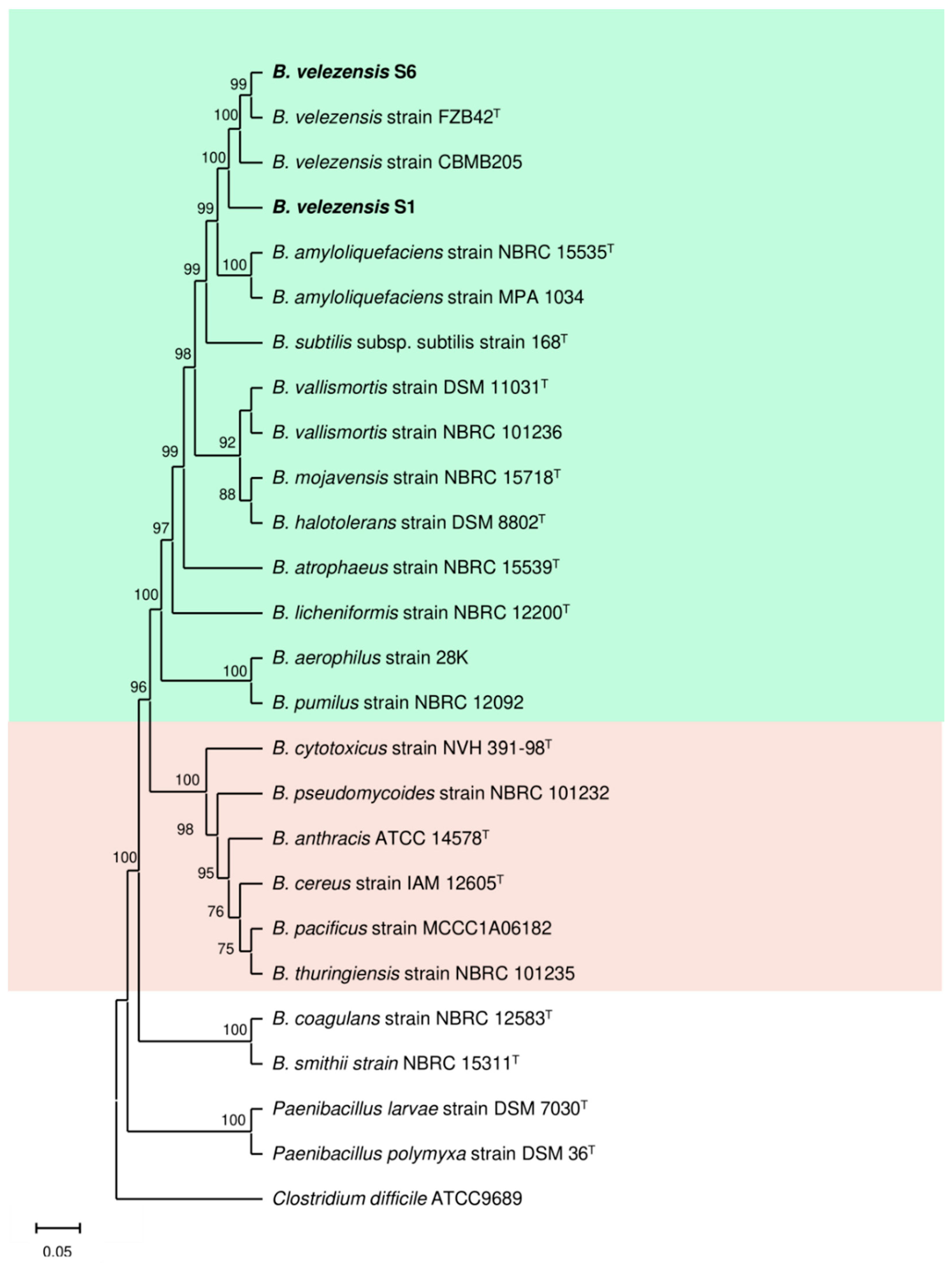
3.1. S1 and S6 Strains Are New Unique B. velezensis Genotypes

Based on the obtained 16S rRNA gene sequences, strains S1 and S6 shared a high sequence similarity (>99%) with B. velezensis. The phylogenetic analysis constructed using MEGA version X with the neighbor-joining method indicated that strains S1 and S6 belong to the genus Bacillus and form a monophyletic cluster with B. velezensis with a high bootstrap score (>99%) (Figure 1). Moreover, the sequence alignment of the 16S rRNA gene sequence between strains S1 and S6 showed that both strains shared 99.86% sequence identity. Therefore, the following names, B. velezensis strain S1 and B. velezensis strain S6, were proposed. They also shared a high sequence identity (>98%) with other species of Bacillus sp., including B. vallismortis, B. subtilis, B. mojavensis, and B. halotolerans (Figure 1).

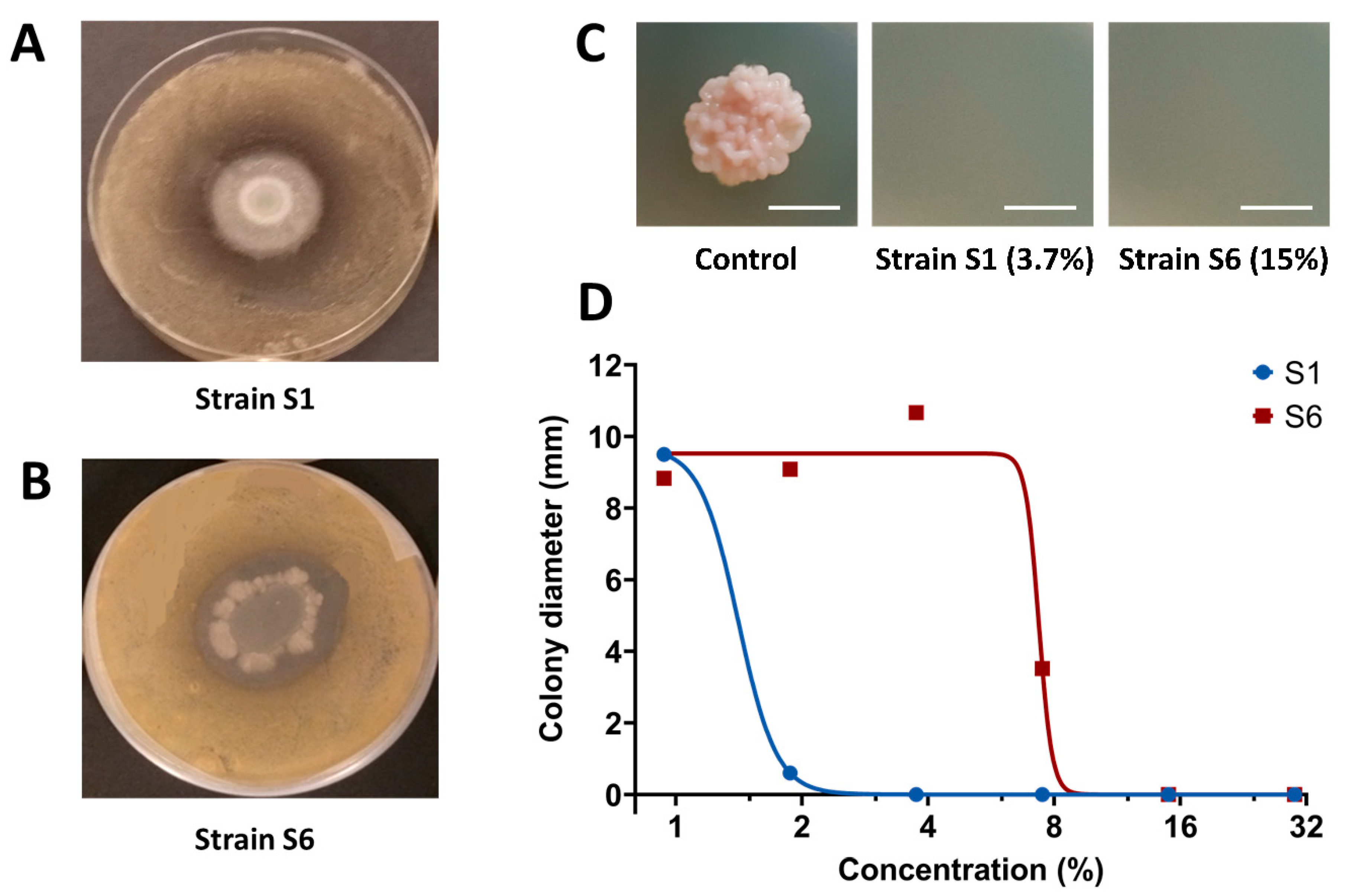
3.2. S1 Strain Shows Stronger Direct In Vitro Antifungal Activity towards Z. tritici Compared to S6 Strain

The antagonistic activity of S1 and S6 strains towards Z. tritici was performed by depositing a bacterial suspension on the center of the PDA-amended Petri plates previously inoculated with Z. tritici. Results revealed that both strains exhibited an antagonistic activity against the pathogen and formed a visible halo around the formed bacterium colonies (Figure 2A,B), therefore revealing a release and diffusion of antimicrobial compounds into the surrounding medium. On the other hand, culture filtrates from the two strains were produced and tested for their antimicrobial activity against the fungus on the PDA medium. Dose-response curves revealed that culture filtrates from both strains were able to inhibit fungal growth, but the S1 culture filtrate showed a higher antifungal activity when compared to the S6 culture filtrate (Figure 2C,D). The minimal-inhibitory dilution (MID) values for the S1 and S6 culture filtrates corresponded to 3.75% and 15%, while the half-maximal inhibitory dilution (ID50) values for the S1 and S6 culture filtrates corresponded to 1.4% and 7.4%, respectively (Figure 2C,D). Lately, and to examine the effect of culture media on the growth of the two bacterial strains for further experiments and optimization, four growth media were tested (King’s B medium, KB; Landy; lysogeny broth, LB; plate count agar, PCA). Results highlighted that LB is the most suitable culture medium, among the four tested ones, for in vitro growth of both strains (Figure S1).

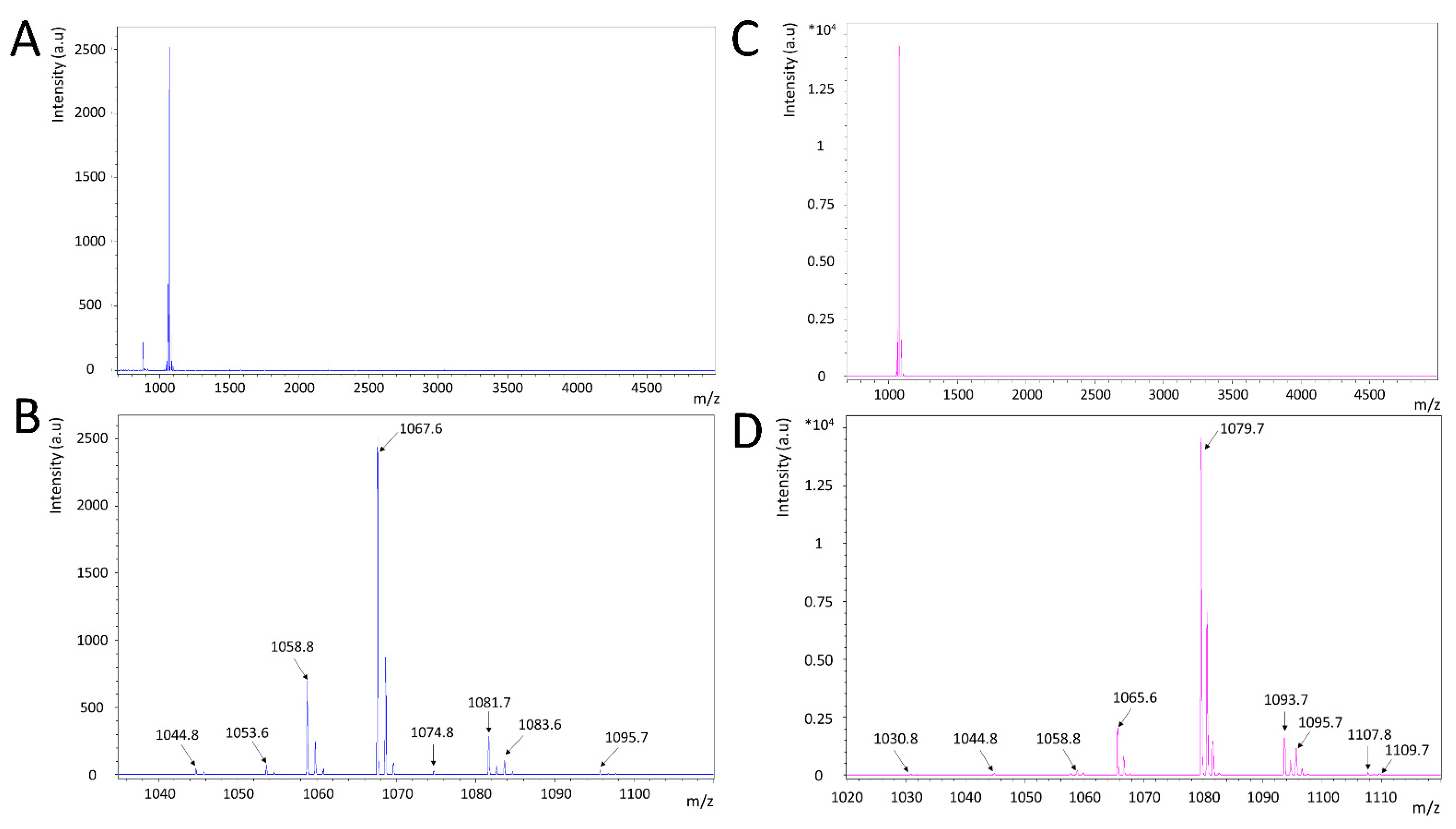
3.3. Both S1 and S6 Strains Produce Cyclic Lipopeptides but with Distinct Patterns

Specialized metabolite production and, in particular, lipopeptide production by the two B. velezensis strains S1 and S6 grown on a solid medium was assessed using MALDI—ToF analysis. Both strains produced molecules with m/z corresponding to cyclic lipopeptides from different families previously described in the Bacillus genera, namely, surfactins, iturins, and fengycins [23] (Table 1, Figure 3). Strains S1 and S6 biosynthesized molecules whose m/z were attributed to analogues of either surfactin or pumilacidin, two lipopeptides of the surfactin family. These analogues would bear various fatty acid tails, i.e., C13 (m/z 1030.8 [M + Na]+), only found in strain S6, C14 (m/z 1044.8 [M + Na]+), and C15 (m/z 1058.8 [M + Na]+, 1074.8 [M + K]+). These m/z could also correspond to many other surfactin and pumilacidin analogues presenting amino acid modifications at different locations on the peptide chain. In the mass range of the iturin family, strain S1 produced molecules whose masses were attributed to several analogues of bacillomycin D, with various fatty acid lengths, i.e., C14 (m/z 1053.6 [M + H]+), C15 (m/z 1067.7 [M + Na]+, 1083.7 [M + K]+), C16 (m/z 1081.7 [M + Na]+), and C17 (m/z 1095.7 [M + Na]+). Strain S6 produced molecules with m/z corresponding to analogues of either iturin A or mycosubtilin. The different analogues would also bear fatty acids of various lengths, i.e., C14 (m/z 1065.6 [M + Na]+), C15 (m/z 1079.6 [M + Na]+, 1095.6 [M + K]+), C16 (m/z 1093.6 [M + Na]+, 1109.6 [M + K]+), and C17 (m/z 1107.6 [M + Na]+. Finally, two peaks that were only detected on the mass spectrum of strain S1 (m/z 1486.0, 1499.8) were putatively assigned to various fengycin A analogues with different possible fatty acid chains (C15 to C17, with or without unsaturation).
Although common lipopeptide masses were detected for the two bacterial strains, they, overall, displayed different lipopeptide production profiles. In particular, strain S1 produces lipopeptides of three different families (surfactins, iturins, fengycins) while strain S6 would produce lipopeptides of only two different families (surfactins and iturins). In addition, iturins produced by strain S1 are putative analogues of bacillomycin D, while those produced by strain S6 would be analogues of either iturin A or mycosubtilin (Table 1).

4. Discussion

We isolated here two wheat phyllosphere-associated bacterial strains identified as two strains of B. velezensis (S1 and S6), with very similar growth abilities in different nutrient media, and exhibiting significant in vitro antagonistic activity against Z. tritici. Our results are in agreement with previous reports showing that this beneficial bacterial species can display antagonistic activity against crop phytopathogens. For instance, the B. velezensis strain FZB42 (previously named B. amyloliquefaciens strain FZB42) has been shown to exhibit in vitro antagonistic activity against another wheat pathogen, Fusarium graminearum [24]. We suggest that the antagonistic activity of the B. velezensis strains S1 and S6 observed here against Z. tritici is due to the capacity of the strains to produce antimicrobial compounds, such as lipopeptides. Indeed, lipopeptides are known to be the major contributor to Bacillus sp. biocontrol activity [25]. Allioui and colleagues [26] have suggested that the biofungicide activity of two Bacillus strains (B. subtilis Alg.24B1 and B. simplex Alg.24B2), isolated in an Algerian field, against Z. tritici may be related to the coproduction of three types of lipopeptides, including surfactins, iturins, and fengycins.
Various strains of B. velezensis have recently received considerable attention, since this species can produce different types of biologically active secondary metabolites that can suppress plant pathogens [27]. It has been reported that B. velezensis strains are able to synthesize various bioactive lipopeptides and polyketides, such as bacillomycin, bacillibactin, fengycin, surfactin, bacillaene, difficidin, and macrolactin [28]. Currently, several products based on Bacillus spp., such as B. amyloliquefaciens, B. pumilus, or B. subtilis, are marketed as biopesticides [27,29].
Our results showed that the culture filtrate from the B. velezensis strain S1 displayed a strong and a higher antifungal activity against Z. tritici when compared to the strain S6. This marked difference could be explained by the differential composition in the lipopeptides synthetized by the two strains. Indeed, MALDI—ToF analysis revealed the presence of fengycin and bacillomycin D (an antimicrobial lipopeptide belonging to the iturin family) only in the most active filtrate, from the strain S1. The importance of these two lipopeptides in the antifungal activity of B. velezensis has already been reported for the B. velezensis strain FZB42. Indeed a double mutant of this strain that was deficient in both bacillomycin D and fengycin synthesis (∆bmyA ∆fenA) was heavily impaired in its ability to inhibit the growth of F. oxysporum, thereby indicating synergistic effects among such lipopeptides against this targeted fungal pathogen [30]. Interestingly, our results suggested that B. velezensis strains S1 and FZB42 shared a high sequence similarity, supporting the hypothesis that fengycin and baccilomycin D are also responsible for strain S1’s antifungal activity. Similarly, Romero and colleagues [8] reported that lipopeptides from the iturin and fengycin families have a major role in the antimicrobial activity of Bacillus sp. strains on Podosphaera fusca. However, other studies suggest that the presence of bacillomycin is solely responsible for the antifungal activity. For instance, bacillomycin D-deficient mutant strains of B. velezensis strain FZB42 (or strain SQR9) exhibited severely impaired antifungal activities, indicating that bacillomycin D appears to be the major contributor to the antifungal activity against F. oxysporum [31,32]. Cao et al. [33] suggest that the relative contribution of each lipopeptide to the antimicrobial activity may also be dependent on the targeted plant pathogen. These authors have reported that iturins and fengycins are functionally redundant in the antagonism against the bacterium Ralstonia solanacearum, when only iturin family metabolites appear to display antagonistic activity against the fungus F. oxysporum.
The effects of purified lipopeptides from B. subtilis have been previously examined against Z. tritici. It has been shown that mycosubtilin, a lipopeptide belonging to the iturin family, displayed a strong antifungal activity against Z. tritici (half-maximal inhibitory concentration of 1.4 mg L−1), while neither surfactin nor fengycin showed significant activity towards the pathogen [34]. On the other hand, bacterium cell-free filtrates of a mutant strain of B. subtilis overproducing mycosubtilin showed an antagonistic activity against several phytopathogenic fungi, including Pythium aphanidermatum, Botrytis cinerea, and F. oxysporum, with growth inhibition zones significantly larger than those induced by the wild-type supernatant [35]. Taken together, these results suggest that lipopeptides of the iturin family may display an interesting antifungal activity towards plant phytopathogens. Moyne et al. [36] reported that, for bacillomycin analogues, variations in their fatty acid length may have an influence on their antifungal activity toward various phytopathogenic fungi, such as Alternaria solani, Aspergillus flavus, Colletotrichum gloeosporioides, Phomopsis gossypii, and Sclerotium rolfsii. The mechanisms underlying bacillomycin D antifungal activity toward F. graminearum were investigated, revealing an inhibition of germination and conidia formation [24].
This study highlighted that the two strains isolated from wheat plant ears and identified as B. velezensis strains S1 and S6 exhibited an in vitro antimicrobial activity against the wheat pathogen Z. tritici, and that the observed biological activity could be related to the production of lipopeptides. The higher antifungal activity observed with the culture filtrate S1 could be due to the different lipopeptides it may produce, in particular the various analogues of bacillomycin D. Hence, further investigations using purified lipopeptides are needed to confirm the antifungal activity of bacillomycin D, alone or in mixture with other lipopeptides, against Z. tritici. On the other hand, Chowdhury et al. [37] suggested that B. velezensis, and notably the strain FBZ42, may trigger pathways of induced systemic resistance (ISR), which can also contribute to the biocontrol activity of FZB42. Indeed, sub-lethal concentrations of lipopeptides and volatiles produced by plant-associated Bacilli trigger pathways of ISR, which protect plants against attacks of pathogenic microbes, viruses, and nematodes. In this sense, the ability of the S1 and S6 B. velezensis strains to induce ISR in wheat against Z. tritici should be further investigated.

Supplementary Materials

The following supporting information can be downloaded at: https://www.mdpi.com/article/10.3390/agronomy12010095/s1, Figure S1: Kinetics of S1 and S6 Bacillus velezensis strains growth in different liquid media. These two strains were inoculated in Erlenmeyer containing one of the four different media, King’s B (KB), Landy, lysogeny broth (LB), and Plate Count Agar (PCA) for three days. Bacterium growth was assessed by measuring the optical density in each well at 600 nm. Three repetitions were used for each condition. Optical density was also measured for controls (i.e., media without bacteria) and these data were used to correct optical density means presented in this figure. Bars stand for standard deviation (n = 3).

Author Contributions

Conceptualization, B.R., E.A.B., C.J., P.H., M.E.G., N.C., S.A., P.T., A.R., N.B., P.R., M.M.-R. and A.S.; methodology, M.E.G., N.C., S.A., N.B., P.T., P.H., M.M.-R., M.S., P.R., Q.E., A.B., A.R. and A.S.; formal analysis, M.E.G., N.C., S.A., A.R., N.B., P.H., M.S., A.B., Q.E., M.M.-R. and R.P.; investigation, A.R., P.H., M.S., R.P., M.M.-R., M.E.G., N.C., Q.E., S.A., A.B. and N.B.; writing—original draft preparation, A.S., M.S. and R.P.; writing—review and editing, M.E.G., N.C., S.A., A.R., N.B., P.H., R.P., Q.E., B.R., P.T., A.B., C.J., E.A.B., P.R., M.M.-R. and A.S.; supervision, B.R., A.R., C.J., E.A.B., P.R., M.M.-R. and A.S.; project administration, P.H., P.R., A.S. and M.M.-R.; funding acquisition, P.H., M.M.-R., P.R. and A.S. All authors have read and agreed to the published version of the manuscript.

Funding

This research was conducted in the framework of the Bacplant project, funded by the European program Arimnet and l’Agence Nation ale de la Recherche (ANR), and the project CPER Alibiotech, funded by the European Union, the French State, and the French Region Hauts-de-France.

Acknowledgments

The authors thank Océane Bueno for technical support.

Conflicts of Interest

The authors declare no conflict of interest. The funders had no role in the design of the study, in the collection, analyses, or interpretation of data, in the writing of the manuscript, or in the decision to publish the results.

References

  1. Bhandari, V.; Ahmod, N.Z.; Shah, H.N.; Gupta, R.S. Molecular Signatures for Bacillus Species: Demarcation of the Bacillus subtilis and Bacillus cereus Clades in Molecular Terms and Proposal to Limit the Placement of New Species into the Genus Bacillus. Int. J. Syst. Evol. Microbiol. 2013, 63, 2712–2726. [Google Scholar] [CrossRef]
  2. Fan, B.; Blom, J.; Klenk, H.-P.; Borriss, R. Bacillus amyloliquefaciens, Bacillus velezensis, and Bacillus siamensis Form an “Operational Group B. amyloliquefaciens” within the B. subtilis Species Complex. Front. Microbiol. 2017, 8, 22. [Google Scholar] [CrossRef] [Green Version]
  3. Hamley, I.W. Lipopeptides: From Self-Assembly to Bioactivity. Chem. Commun. 2015, 51, 8574–8583. [Google Scholar] [CrossRef] [Green Version]
  4. Strieker, M.; Tanović, A.; Marahiel, M.A. Nonribosomal Peptide Synthetases: Structures and Dynamics. Curr. Opin. Struct. Biol. 2010, 20, 234–240. [Google Scholar] [CrossRef]
  5. Deravel, J.; Lemière, S.; Coutte, F.; Krier, F.; Van Hese, N.; Béchet, M.; Sourdeau, N.; Höfte, M.; Leprêtre, A.; Jacques, P. Mycosubtilin and Surfactin Are Efficient, Low Ecotoxicity Molecules for the Biocontrol of Lettuce Downy Mildew. Appl. Microbiol. Biotechnol. 2014, 98, 6255–6264. [Google Scholar] [CrossRef]
  6. Xun-Chao, C.; Hui, L.; Ya-Rong, X.; Chang-Hong, L. Study of Endophytic Bacillus amyloliquefaciens CC09 and Its Antifungal Cyclic Lipopeptides. J. Appl. Biol. Biotechnol. 2013, 1, 1–5. [Google Scholar] [CrossRef]
  7. Guo, Q.; Dong, W.; Li, S.; Lu, X.; Wang, P.; Zhang, X.; Wang, Y.; Ma, P. Fengycin Produced by Bacillus subtilis NCD-2 Plays a Major Role in Biocontrol of Cotton Seedling Damping-off Disease. Microbiol. Res. 2014, 169, 533–540. [Google Scholar] [CrossRef]
  8. Romero, D.; de Vicente, A.; Rakotoaly, R.H.; Dufour, S.E.; Veening, J.-W.; Arrebola, E.; Cazorla, F.M.; Kuipers, O.P.; Paquot, M.; Pérez-García, A. The Iturin and Fengycin Families of Lipopeptides Are Key Factors in Antagonism of Bacillus subtilis toward Podosphaera fusca. Mol. Plant Microbe Interact. 2007, 20, 430–440. [Google Scholar] [CrossRef] [Green Version]
  9. Jacques, P. Surfactin and Other Lipopeptides from Bacillus spp. In Biosurfactants: From Genes to Applications; Soberón-Chávez, G., Ed.; Microbiology Monographs; Springer: Berlin/Heidelberg, Germany, 2011; pp. 57–91. ISBN 978-3-642-14490-5. [Google Scholar]
  10. Crouzet, J.; Arguelles-Arias, A.; Dhondt-Cordelier, S.; Cordelier, S.; Pršić, J.; Hoff, G.; Mazeyrat-Gourbeyre, F.; Baillieul, F.; Clément, C.; Ongena, M.; et al. Biosurfactants in Plant Protection Against Diseases: Rhamnolipids and Lipopeptides Case Study. Front. Bioeng. Biotechnol. 2020, 8. [Google Scholar] [CrossRef]
  11. Fira, D.; Dimkić, I.; Berić, T.; Lozo, J.; Stanković, S. Biological Control of Plant Pathogens by Bacillus Species. J. Biotechnol. 2018, 285, 44–55. [Google Scholar] [CrossRef]
  12. Ponomarenko, A.; Goodwin, S.B.; Kema, G.H.J. Septoria Tritici Blotch (STB) of Wheat. In Plant Health Instructor; American Phytopathological Society: St. Paul, MI, USA; p. 2011. [CrossRef]
  13. Fones, H.; Gurr, S. The Impact of Septoria Tritici Blotch Disease on Wheat: An EU Perspective. Fungal Genet. Biol. 2015, 79, 3–7. [Google Scholar] [CrossRef] [Green Version]
  14. Siah, A.; Deweer, C.; Duyme, F.; Sanssené, J.; Durand, R.; Halama, P.; Reignault, P. Correlation of in Planta Endo-Beta-1,4-Xylanase Activity with the Necrotrophic Phase of the Hemibiotrophic Fungus Mycosphaerella graminicola. Plant Pathol. 2010, 59, 661–670. [Google Scholar] [CrossRef]
  15. Lovell, D.J.; Hunter, T.; Powers, S.J.; Parker, S.R.; Bosch, F.V. den Effect of Temperature on Latent Period of Septoria Leaf Blotch on Winter Wheat under Outdoor Conditions. Plant Pathol. 2004, 53, 170–181. [Google Scholar] [CrossRef]
  16. Cowger, C.; Hoffer, M.E.; Mundt, C.C. Specific Adaptation by Mycosphaerella graminicola to a Resistant Wheat Cultivar. Plant Pathol. 2000, 49, 445–451. [Google Scholar] [CrossRef]
  17. Cheval, P.; Siah, A.; Bomble, M.; Popper, A.D.; Reignault, P.; Halama, P. Evolution of QoI Resistance of the Wheat Pathogen Zymoseptoria tritici in Northern France. Crop Prot. 2017, 92, 131–133. [Google Scholar] [CrossRef]
  18. Beneduzi, A.; Ambrosini, A.; Passaglia, L.M.P. Plant Growth-Promoting Rhizobacteria (PGPR): Their Potential as Antagonists and Biocontrol Agents. Genet. Mol. Biol. 2012, 35, 1044–1051. [Google Scholar] [CrossRef] [Green Version]
  19. Landy, M.; Warren, G.H.; RosenmanM, S.B.; Colio, L.G. Bacillomycin: An Antibiotic from Bacillus subtilis Active against Pathogenic Fungi. Proc. Soc. Exp. Biol. Med. 1948, 67, 539–541. [Google Scholar] [CrossRef]
  20. Esmaeel, Q.; Jacquard, C.; Sanchez, L.; Clément, C.; Ait Barka, E. The Mode of Action of Plant Associated Burkholderia against Grey Mould Disease in Grapevine Revealed through Traits and Genomic Analyses. Sci Rep. 2020, 10, 19393. [Google Scholar] [CrossRef]
  21. Kumar, S.; Stecher, G.; Li, M.; Knyaz, C.; Tamura, K. MEGA X: Molecular Evolutionary Genetics Analysis across Computing Platforms. Mol. Biol. Evol. 2018, 35, 1547–1549. [Google Scholar] [CrossRef]
  22. Platel, R.; Chaveriat, L.; Le Guenic, S.; Pipeleers, R.; Magnin-Robert, M.; Randoux, B.; Trapet, P.; Lequart, V.; Joly, N.; Halama, P.; et al. Importance of the C12 Carbon Chain in the Biological Activity of Rhamnolipids Conferring Protection in Wheat against Zymoseptoria tritici. Molecules 2021, 26, 40. [Google Scholar] [CrossRef]
  23. Ongena, M.; Jacques, P. Bacillus Lipopeptides: Versatile Weapons for Plant Disease Biocontrol. Trends Microbiol. 2008, 16, 115–125. [Google Scholar] [CrossRef]
  24. Gu, Q.; Yang, Y.; Yuan, Q.; Shi, G.; Wu, L.; Lou, Z.; Huo, R.; Wu, H.; Borriss, R.; Gao, X. Bacillomycin D Produced by Bacillus amyloliquefaciens Is Involved in the Antagonistic Interaction with the Plant-Pathogenic Fungus Fusarium graminearum. Appl. Environ. Microbiol. 2017, 83. [Google Scholar] [CrossRef] [Green Version]
  25. Abderrahmani, A.; Tapi, A.; Nateche, F.; Chollet, M.; Leclère, V.; Wathelet, B.; Hacene, H.; Jacques, P. Bioinformatics and Molecular Approaches to Detect NRPS Genes Involved in the Biosynthesis of Kurstakin from Bacillus thuringiensis. Appl. Microbiol. Biotechnol. 2011, 92, 571–581. [Google Scholar] [CrossRef]
  26. Allioui, N.; Driss, F.; Dhouib, H.; Jlail, L.; Tounsi, S.; Frikha-Gargouri, O. Two Novel Bacillus Strains (Subtilis and Simplex Species) with Promising Potential for the Biocontrol of Zymoseptoria tritici, the Causal Agent of Septoria Tritici Blotch of Wheat. BioMed Res. Int. 2021, 2021, 6611657. [Google Scholar] [CrossRef]
  27. Rabbee, M.F.; Ali, M.S.; Choi, J.; Hwang, B.S.; Jeong, S.C.; Baek, K. Bacillus velezensis: A Valuable Member of Bioactive Molecules within Plant Microbiomes. Molecules 2019, 24, 1046. [Google Scholar] [CrossRef] [Green Version]
  28. Chen, J.; Wu, Q.; Hua, Y.; Chen, J.; Zhang, H.; Wang, H. Potential Applications of Biosurfactant Rhamnolipids in Agriculture and Biomedicine. Appl. Microbiol. Biotechnol. 2017, 101, 8309–8319. [Google Scholar] [CrossRef]
  29. Pérez-García, A.; Romero, D.; de Vicente, A. Plant Protection and Growth Stimulation by Microorganisms: Biotechnological Applications of Bacilli in Agriculture. Curr. Opin. Biotechnol. 2011, 22, 187–193. [Google Scholar] [CrossRef]
  30. Koumoutsi, A.; Chen, X.-H.; Henne, A.; Liesegang, H.; Hitzeroth, G.; Franke, P.; Vater, J.; Borriss, R. Structural and Functional Characterization of Gene Clusters Directing Nonribosomal Synthesis of Bioactive Cyclic Lipopeptides in Bacillus amyloliquefaciens Strain FZB42. J. Bacteriol. 2004, 186, 1084–1096. [Google Scholar] [CrossRef] [Green Version]
  31. Chen, X.H.; Koumoutsi, A.; Scholz, R.; Schneider, K.; Vater, J.; Süssmuth, R.; Piel, J.; Borriss, R. Genome Analysis of Bacillus amyloliquefaciens FZB42 Reveals Its Potential for Biocontrol of Plant Pathogens. J. Biotechnol. 2009, 140, 27–37. [Google Scholar] [CrossRef]
  32. Xu, Z.; Shao, J.; Li, B.; Yan, X.; Shen, Q.; Zhang, R. Contribution of Bacillomycin D in Bacillus amyloliquefaciens SQR9 to Antifungal Activity and Biofilm Formation. Appl. Environ. Microbiol. 2013, 79, 808–815. [Google Scholar] [CrossRef] [Green Version]
  33. Cao, Y.; Pi, H.; Chandrangsu, P.; Li, Y.; Wang, Y.; Zhou, H.; Xiong, H.; Helmann, J.D.; Cai, Y. Antagonism of Two Plant-Growth Promoting Bacillus velezensis Isolates Against Ralstonia solanacearum and Fusarium oxysporum. Sci. Rep. 2018, 8, 4360. [Google Scholar] [CrossRef]
  34. Mejri, S.; Siah, A.; Coutte, F.; Magnin-Robert, M.; Randoux, B.; Tisserant, B.; Krier, F.; Jacques, P.; Reignault, P.; Halama, P. Biocontrol of the Wheat Pathogen Zymoseptoria tritici Using Cyclic Lipopeptides from Bacillus subtilis. Environ. Sci. Pollut. Res. 2018, 25, 29822–29833. [Google Scholar] [CrossRef]
  35. Leclère, V.; Béchet, M.; Adam, A.; Guez, J.-S.; Wathelet, B.; Ongena, M.; Thonart, P.; Gancel, F.; Chollet-Imbert, M.; Jacques, P. Mycosubtilin Overproduction by Bacillus subtilis BBG100 Enhances the Organism’s Antagonistic and Biocontrol Activities. Appl. Environ. Microbiol. 2005, 71, 4577–4584. [Google Scholar] [CrossRef] [Green Version]
  36. Moyne, A.L.; Shelby, R.; Cleveland, T.E.; Tuzun, S. Bacillomycin D: An Iturin with Antifungal Activity against Aspergillus flavus. J. Appl. Microbiol. 2001, 90, 622–629. [Google Scholar] [CrossRef]
  37. Chowdhury, S.P.; Hartmann, A.; Gao, X.; Borriss, R. Biocontrol Mechanism by Root-Associated Bacillus amyloliquefaciens FZB42—A Review. Front. Microbiol. 2015, 6, 780. [Google Scholar] [CrossRef] [Green Version]
Figure 1. Phylogenetic tree of the S1 and S6 Bacillus velezensis strains. The tree was built based on 16S rRNA gene sequences using MEGA version X with the neighbor-joining method. Values at the nodes indicate bootstrap values out of 1000 resamplings. Green color = members of the ‘Subtilis group’ of Bacillus species, Pink = members of the ‘Cereus group’, colorless = other members of Bacillus and Paenibacillus species. Clostridium difficile ATCC9689 was used as an outgroup. S1 and S6 are shown in bold. Type strains are indicated as (T).
Agronomy 12 00095 g001
Figure 2. Antifungal activity of S1 and S6 Bacillus velezensis strains against the Zymoseptoria tritici strain T02596. (A,B), Antagonistic activity of S1 (A) and S6 (B) strains on solid PDA medium at 13 days after Petri plate inoculation. The inhibiting effect is visualized by the formation of a halo around the bacterial colonies. (C) Illustration of the antifungal effect of the S1 and S6 culture filtrates on Z. tritici observed on PDA medium. (D) In vitro dose-response curves of the antifungal effect of the S1 and S6 culture filtrates on Z. tritici growth, assessed by measuring the fungal colony diameters at 10 days after plate inoculation. Dose-response curves were performed using GraphPad Prism software v 9.0. Three repetitions were used as replicates for each concentration of tested culture filtrate. Scale-bars stand for 5 mm.
Agronomy 12 00095 g002
Figure 3. Whole-cell MALDI—ToF mass spectra of the two Bacillus velezensis strains S1 (A,B) and S6 (C,D). (B,D) are zooms on (A,C), respectively.
Agronomy 12 00095 g003
Table 1. Putative metabolites produced by the two Bacillus velezensis bacterial strains S1 and S6 grown on PDA medium, detected by MALDI—TOF.
Bacillus velezensis S1
m/zPutative Assigned LipopeptideLipopeptide Family
1044.8Surfactin C14 or [Val7] Pumilacidin C14 [M + Na]+Surfactin
1058.8Surfactin C15 or [Val7] Pumilacidin C15 [M + Na]+Surfactin
1074.8Surfactin C15 or [Val7] Pumilacidin C15 [M + K]+Surfactin
1053.6Bacillomycin D C14 [M + Na]+Iturin
1067.7Bacillomycin D C15 [M + Na]+Iturin
1083.7Bacillomycin D C15 [M + K]+Iturin
1081.7Bacillomycin D C16 [M + Na]+Iturin
1095.7Bacillomycin D C17 [M + Na]+Iturin
1486.0Fengycin A C16 [M + Na]+ or Fengycin A C15 (insat) [M + K]+Fengycin
1499.8Fengycin A C17 [M + Na]+ or Fengycin AC16 (insat) [M + K]+ [M + Na]+Fengycin
Bacillus velezensis S6
m/zPutative Assigned LipopeptideLipopeptide Family
1030.8Surfactin C13 or [Val7] Pumilacidin C13 [M + Na]+Surfactin
1044.8Surfactin C14 or [Val7] Pumilacidin C14 [M + Na]+Surfactin
1058.8Surfactin C15 or [Val7] Pumilacidin C15 [M + Na]+Surfactin
1065.6Iturin A C14 or Mycosubtilin C14 [M + Na]+Iturin
1079.6Iturin A C15 or Mycosubtilin C15 [M + Na]+Iturin
1095.6Iturin A C15 or Mycosubtilin C15 [M + K]+Iturin
1093.6Iturin A C16 or Mycosubtilin C16 [M + Na]+Iturin
1109.6Iturin A C16 or Mycosubtilin C16 [M + K]+Iturin
1107.6Iturin A C17 or Mycosubtilin C17 [M + Na]+Iturin
Publisher’s Note: MDPI stays neutral with regard to jurisdictional claims in published maps and institutional affiliations.

