Article

Phytotoxicity and Plant Defence Induction by Cinnamomum cassia Essential Oil Application on Malus domestica Tree: A Molecular Approach

by
Pierre-Yves Werrie
1,*,
Anthony Juillard
2,
Christelle Heintz
2,
Marie-Noëlle Brisset
2 and
Marie-Laure Fauconnier
1
1
Laboratory of Chemistry of Natural Molecules, Gembloux Agro-Bio Tech, University of Liège, B-5030 Gembloux, Belgium
2
University Angers, Institut Agro, INRAE, IRHS, SFR QUASAV, F-49000 Angers, France
*
Author to whom correspondence should be addressed.
Agronomy 2022, 12(2), 512; https://doi.org/10.3390/agronomy12020512
Submission received: 11 January 2022 / Revised: 14 February 2022 / Accepted: 17 February 2022 / Published: 18 February 2022

Abstract

:
Essential oils (EOs) are actively investigated as an alternative to numerous synthetic biocide products. Due to their large spectra of biological activities, the impact of EOs on non-target organisms should be characterized for biopesticide development purposes. In this study the potential phytotoxicity of Cinnamomum cassia EO (CEO) on apple trees (Malus domestica) was investigated in terms of oxidative burst (glutathione redox state) and damage (malondialdehyde). At 2%, CEO concentration the reduced glutathione leaf content drops from 269.6 ± 45.8 to 143 ± 28.4 nmol g−1FW, after 30 min, illustrating a rapid and strong oxidative burst. Regarding oxidative damage, malondialdehyde increased significantly 24 h post application to 10.7 ± 3.05 nmol g−1FW. Plant defence induction was previously suspected after trans-cinnamaldehyde (CEO main compound) application. Therefore, the elicitor potential was investigated by qRT-PCR, on the expression level of 29 genes related to major defence pathways (PR protein, secondary metabolism, oxidative stress, parietal modification). Multivariate analysis and increased expression levels suggest induction of systemic resistance. Hence, the present research illustrates the dose–dependent phytotoxicity of CEO in terms of lipid peroxidation. Transcriptional data illustrates the elicitor properties of CEO. These findings can help to design pest management strategies considering both their risks (phytotoxicity) and benefits (defence activation combined with direct biocide properties).

1. Introduction

Owing to their antibacterial [1], fungicidal [2], insecticidal [3], acaricidal [4], nematicidal [5] and herbicidal [6] properties, essential oils (EO) are increasingly investigated to be included in agricultural practices as biopesticides. According to Dayan et al. [7], the fungicidal mode of action (Moa) consists of the inhibition of synthesis of the fungal cell wall component chitin. In addition, EOs have some properties that make them suitable for insects’ management. EO physiological actions on insects suggest a neurotoxic Moa [8,9], notably through octopamine synapses, γ-aminobutyric acid (GABA) and acetylcholinesterase (AChE) inhibition [10,11]. Recent studies reveal larger impacts on the development and functioning of the muscular and nervous systems, cellular respiration, protein synthesis, and detoxification [12]. The aforementioned biological properties make them a good alternative to synthetic pesticides. Moreover, they follow the European directive (2009) of a reduced risk for human health and to the environment. Their phytotoxic properties make them suitable for weed control, but are not desirable in other application contexts. In order to enable their large-scale use in the field, their potential phytotoxicity with non-target organisms, especially crop plants from an agronomic perspective must be assayed.
Adverse physiological impacts following EO application are disparate: water status alteration, inhibition of respiration and photosynthesis, membrane interaction/disruption, reactive oxygen/nitrogen species induction, microtubule disruption and enzymatic or phytohormones regulation [13]. From a mechanistic point of view, most of these alterations originate from, or lead to, reactive oxygen species (ROS) production. An oxidative burst following abiotic stresses is one of the largest shared responses in plants. The cell redox state modification, if not properly handled by the antioxidant system, can result in oxidative damage and lead to programmed cell death (PCD). Therefore, ROS were long considered as a toxic by-product of metabolism. Nevertheless, they also play a key role as regulators of growth and defence pathways [14]. Plant cells are well equipped to efficiently scavenge ROS and their reaction products by the coordinated action of non-enzymatic and enzymatic antioxidant components. Among the non-enzymatic ones, glutathione is a major component of the ascorbate–glutathione (AsA–GSH) pathway, playing a significant role in protecting cells against ROS-accrued potential anomalies [15]. Most data suggest that enhanced ROS availability, especially hydrogen peroxide (H2O2), has less impact on the ascorbate–dehydroascorbate (DHA) ratio than on the redox status of the glutathione pool [16]. Various stress conditions drive characteristic changes in the intracellular amount and redox state of glutathione. Thus, modifications in the whole glutathione status can be taken as a reliable marker of the degree of intracellular oxidative stress [17,18]. The main function of glutathione consists of a redox-homeostatic buffering, serving as a ROS scavenger, but it also plays a role in stress perception, signalling and defence reactions [19,20].
In biological systems, oxygen-derived free radicals have repeatedly been demonstrated to play a role in cellular injury through chain reactions leading to the degradation of macromolecules such as lipids, carbohydrates, proteins and DNA [21]. Indeed, much of the injury caused by exposure to biotic and abiotic stresses is associated with oxidative damage at the cellular level, particularly losses in bio-membrane integrity due to formation of lipid peroxides [22]. It should be noted that following a pathogen invasion or injury, this reaction may also originate from increased lipoxygenase activity [23]. Primary lipid hydroperoxides are highly unstable and reactive, quantification of lipid peroxidation is usually estimated by focusing on secondary oxidation products derived from them [24], such as malondialdehyde (MDA). In studies related to oxidative stress, the measurement of MDA content has been demonstrated to be a reliable lipid peroxidation marker, representative of a rather late stage of oxidation [23,25]. The accumulation of MDA following EO application is frequently observed with, for example, Origanum vulgare [26], Artemisia Fragrans [27], Cymbopogon citratus [28] or pure compounds including cinnamaldehyde [29].
After treatment with EOs, a decrease in the photosynthetic pigments namely chlorophylls and carotenoids in a dose-dependent way have also been reported, resulting from a direct pigment degradation or from an impairment in pigment biosynthetic pathways [28,30]. Total leaf chlorophyll (Chl) content is a popular trait used to get an idea of the plant’s photosynthetic capacity. Chl a and Chl b are the two forms of pigments that predominate in higher plants. Differently involved in light assimilation, Chl a is linked to the photosystems energy-processing centres whereas Chl b is an accessory pigment for harvesting light energy and transmitting it to Chl a [31]. Concerning carotenoids (Car), they act first as collectors of light energy driving photosynthetic processes. As antioxidants, their second role is the protection of the photosynthetic system against detrimental effects of light and O2 (photo-oxidation) by scavenging ROS and the quenching of Chl excited states [32,33].
Apart from phytotoxicity, glutathione and malondialdehyde play a role as regulators of plant defence pathways [34,35,36]. Moreover, monoterpenoids are able to activate defence genes by signalling processes and Ca2+ influx causes by membrane depolarization, protein phosphorylation/dephosphorylation and the action of ROS [37]. This gene expression can either lead to priming (an accelerated gene-response to biotic stress) or direct defence elicitations. Priming properties have been observed in wheat seed with application of thyme EO [38] and also in apple against Botrytis cinerea with thyme and savory EO [39]. Priming following exposure to mint volatiles resulted in enhanced transcript levels of defence genes in soy through histone acetylation within the promoter regions [40]. Regarding defence elicitation, systematic resistance induction is divided between systemic acquired resistance (SAR) and induced systemic resistance (ISR). A complex crosstalk exists between the two systems relying on salicylic acid (SA) and jasmonic acid (JA) hormones. Transcriptomic studies following exposure to volatile monoterpenes myrcene and ocimene demonstrated that plants develop a similar response to that induced by methyl jasmonate (MeJA) [41]. The induction of SAR by EO has also been acknowledged in multiple pathosystems with nerodiol on tea plant [42], thyme [43] and clove EO [44] on tomato, cinnamomum zeylanicum and trans-cinnamaldehyde EO on tangerine [45], and citronellal on coffee plant [46]. Expression of defence-related genes is considered the hallmark to decipher the potential elicitor properties [47].
For example, Cinnamomum cassia EO (CEO) has been previously commercialized by the Mycotech Corporation U.S. company as an aphicide/miticide/fungicide based on cinnamaldehyde (30% in the formulation) as the active ingredient [48]. Cinnamaldehyde is also synthesized chemically for use as a fungicide in agriculture (e.g., VertigoTM, CinnacureTM) on a variety of crops. Depending on the biological activity targeted, different concentrations of EO can be applied. Indeed, in vivo herbicidal activity of cinnamaldehyde has been observed at 3% (v/v) concentration on A. thaliana leaves [49]. Field insecticidal activity against two spotted mites in cherry fruit was observed after five applications at 0.25% (v/v) concentration [50]. C. zeylanicum and cinnamaldehyde was applied against Alternaria brown spot in tangerines in the field at 0.1% (v/v) [45]. Finally, EPA registration for CinnacureTM has recommended an application rate at 0.4% (v/v) as a fungicide and insecticide in fruit trees [51].
The objective of this study is to investigate the molecular mechanisms resulting from different concentrations of Cinnamomum cassia EO application (1–2%) on young Malus domestica trees, especially the resulting oxidative burst and the potential oxidative damage. Moreover, the potential plant gene defence activation properties have been investigated by following 29 transcripts from major defence pathways.

2. Materials and Methods

2.1. Plant Material

Experiments on redox status, oxidative damage and photosynthetic pigments were conducted on mature leaves of two-year-old micropropagated Malus domestica Borkh (var. Jonagold) apple trees (height = 53 ± 8 cm; diameter = 4.4 ± 0.6 mm). They were kept in a climate chamber under the following conditions: 21 ± 0.5 °C, 60 ± 10% RH, 16:8 h light: dark periods and a photosynthetic active radiation (PAR) intensity equal to 50 µmol/m2s.
Experiments for transcriptional studies were conducted on open-pollinated apple seedlings (4–6 leaves) of cv. Golden Delicious, grown under greenhouse conditions (natural photoperiod supplemented with artificial light if needed, 17 °C night and 20–23 °C day according to the sun light).

2.2. Emulsion Formulation and Application

A cinnamon EO emulsion (1–2% (v/v) Pranarôm, batch number: CCB114) was obtained using Tween 80 (2%). The emulsion was stabilized using high speed homogenization (HSH) at 9500 rpm for 6 min (Ultra-Turrax T25) followed by high pressure homogenization (HPH) with 8 cycles at 5000 psi (FMC). Following a previously published protocol [47,52,53,54] approximately 30 mL of solution was applied on each plot to runoff.

2.3. Redox Status: Determination of Reduced (GSH) and Oxidized Glutathione (GSSG)

GSH can be derivated using monobromobimane (MBB), the amount of GSH–MBB adduct formed was then measured by high performance liquid chromatography with fluorescence detection (HPLC–FLD). As MBB only reacts with the reduced form GSH, the content of oxidized glutathione GSSG in the samples must be reduced by the addition of dithiothreitol (DTT) in order to obtain the total amount of glutathione (i.e. GSH + GSSG). In this way, the approximate redox state of glutathione can be estimated. The developed method is based on [17,18,55]. Briefly described, apple leaves were ground in liquid nitrogen. 100 mg of this powder was mixed with 1 mL of ice-cold acid extraction buffer (0.4 M HCl, 1 mM Na2EDTA, 1% PVP). The samples were vortexed, centrifuged at 12,000 rpm for 2 min and the supernatant was filtered through a 0.45 µm syringe filter. For GSH 100 µL of supernatant was mixed with 100 µL NaHCO3, 20 µL H2O, 200 µL CHES (0.5M, pH 9) and 20 µL MBB (30 Mm in acetonitrile) and left to incubate for 15 min in the dark on ice. The reaction was then stopped by adding 660 µL of acetic acid (10%), followed by transfer to amber vials. For GSH + GSSG 100 µL of supernatant was neutralized and reduced in Eppendorfs with addition of 100 µL NaHCO3 + 20 µL DTT and incubated for 30 min in the dark and on ice. Then 200 µL CHES and 20 µL MBB were added. The reaction medium was left to incubate for 15 min in the dark and on ice. The reaction was then stopped by adding 660 µL of acetic acid (10%), followed by transfer to amber vials. All analyses were performed on an Agilent 1260 Infinity HPLC system equipped with a FLD detector (λex: 395 nm, λem: 477 nm). The autosampler was thermostated at 6 °C and 50 µL was injected onto the Eclipse XDB-C18 column (150 × 4.6mm, 5µm). The GSH-bimane derivatives were separated from the other molecules using a linear gradient of 0.25% (v/v) acetic acid (pH 3.5) as solvent A, and 100% methanol as solvent B, at a flow rate of 0.8 mL min−1 and a column temperature of 40 °C. The linear gradient started at 18% (v/v) solvent B until 17.5 min then increased to 100% (v/v) solvent B from 20 min to 27.5 min and returned to original condition 18% (v/v) solvent B at 28 min until the run ended at 32.5 min. GSH typical sample chromatogram, calibration curve, LOD and LOQ are available on Figure S1.

2.4. Oxidative Damage

2.4.1. Determination of Malondialdehyde (MDA)

The thiobarbituric acid reactive substances (TBARS) content was determined according to the method of [24] with modifications mainly based on [31,56]. Apple leaves were ground in liquid nitrogen with a mortar and pestle. 100 mg of this powder was mixed in an Eppendorf with 1 mL of ice-cold 5% (w/v) HCl. The samples were vortexed and centrifuged at 13,400 rpm for 10 min. 200 µL of supernatant was added to 40 µL of BHT (0.1% EtOH) and 760 µL of TBA (0.5% in MPA 20%), giving a final pH of approximately 1.0. The reaction mixture was heated for 30 min at 95 °C and then quickly cooled on ice. Once the reaction had stopped the reaction mixture was centrifuged at 4000× g for 5 min and the supernatant was placed in a vial. All analyses were performed on an Agilent 1200 series HPLC system with MWD detector (RF-10AXL). Chromatograms were monitored at 532 nm and the injection volume was 10 µL. Samples were analysed on a Halo® C18 75 × 4.6 mm, 2.7 µm column thermostated at 40 °C and eluted isocratically with 35% MeOH in 50 mM KPO4 buffer (pH 6.8) at 1 mL min−1. MDA typical sample chromatogram, calibration curve, LOD and LOQ are available on Figure S2.

2.4.2. Determination of Photosynthetic Pigments

50 mg of leaf sample were ground in liquid nitrogen with a mortar and pestle. After 15 min extraction on ice in the dark in 10 mL of 96% (v/v) ethanol, the extract was centrifuged at 4000 rpm for 10 min at 4 °C. The absorbance of the supernatant was measured at 470, 649 and 665 nm using an Ultrospec 7000 spectrophotometer.
The concentrations of chlorophyll a and b and carotenoids were calculated as follows:
Ca (μg/g FW) = [(13.36 * A665) − (5.19 * A649)]/sample mass.
Cb (μg/g FW) = [(27.43 * A649) − (8.12 * A665)]/sample mass.
Ccarotenoids (μg/g FW) = [(1000*A470 − 2.13*Ca − 97.64*Cb)/209]/sample mass.

2.5. Induction of Defences (RT-PCR)

At each sampling time (24, 48 and 72 h), the five youngest expanded leaves per modality were collected, pooled, frozen in liquid nitrogen, and stored at −80 °C until extraction. Each experiment was repeated four times. RNA extraction, reverse transcription and real-time quantitative PCR were performed as previously described [57] using the same proprietary primer set for the 29 defence genes and 3 reference genes [58]. Relative changes in defence genes’ expression (log2 ratio) were calculated using the 2-ΔΔCT method with 3 internal reference genes’ for normalization, and against initial time (T0) from control plants

2.6. Data Analysis

All data were collected in Excel and processed using R studio software (version 4.1.2), with all results presented as a boxplot using the ggplot2 package. The main statistical procedure performed was a simple two-factor analysis of variance (ANOVA 2) the fixed factors were time and treatment. The samples came from a randomized design which guarantees their independence. Normality was assayed by the Shapiro–Wilk normality test. Homogeneity of variance was also demonstrated by Levene’s test. In case of interaction, ANOVA tests were performed at each time point independently, followed by a pairwise analysis (t-test). A probability cut-off of α = 0.05, was used for tests of significance in all statistical analyses and adjusted with the Bonferroni correction. As qRT–PCR data were non-normally distributed, the nonparametric Kruskall–Wallis test was applied followed by the Conover post-hoc test with holm correction. Multivariate visualization was performed with heatmap and principal component analysis (PCA) using Complex Heatmap and FactoMiner packages.

3. Results

3.1. Redox Status: Determination of Reduced (GSH) and Oxidized Glutathione (GSSG)

The soluble tripeptide GSH (L-γ-glutamyl-L-cysteinyl-glycine) is the principal low-molecular-weight thiol compound in plants [22]. Glutathione typically accumulates in plant tissues in the range of 200–600 nmol g−1FW [16]. GSH was derivated using monobromobimane (MBB), the amount of GSH-MBB adduct formed was then quantified by HPLC-FLD. As MBB only reacts with the reduced form GSH, the content of oxidized glutathione GSSG in the samples must be reduced by the addition of dithiothreitol (DTT) in order to obtain the total amount of glutathione (i.e., GSH+GSSG).
The results presented in Figure 1 display the reduced glutathione (GSH) leaf content, total content (GSH+GSSG) and GSH/(GSH+GSSG) ratio, over time, after CEO treatment at two concentrations, or tween. Those contents are between 100.7 nmol g−1FW and 486.6 nmol g−1FW. The relatively large standard deviation in the boxplot highlight the heterogeneity of glutathione content between and within Malus domestica leaves. The two-way ANOVA displayed on the top of the graph present significant interactions between the treatment and time implying that the treatment effect is time dependent. This result is consistent with the transitory aspect of the oxidative burst and with the existence of circadian variation within the glutathione ascorbate cycle [59]. However pairwise t-test comparisons at each time shows that the GSH content, as well as its ratio, is significantly decreased after 30 min following 2% CEO applications. This result underlines indirectly, the production of ROS i.e., the oxidative burst occurring rapidly after CEO application. Under normal (unstressed) conditions, it is maintained mostly in its reduced form, resulting in a GSH/GSSG ratio of 10 to 1 (i.e., GSH/(GSH + GSSG) = 91%) [46]. In contrast, under oxidative conditions, two GSH molecules react together to form glutathione disulfide (GSSG) [47]. The specific enzyme glutathione reductase (GR), reduces GSSG back to GSH [14]. Therefore, GSH fluctuates in cells between two different forms: reduced GSH and oxidized GSSG, as a function of GR activity (with NADPH as an electron donor) [48]. The proportion of GSSG increases substantially only in a strongly oxidizing environment [14]. Therefore, a decrease in GSH and the GSH/(GSH + GSSG) ratio is interpreted as evidence of redox imbalance [47]. Indeed, it was previously established that detoxification of H2O2 through the glutathione–ascorbate cycle leads to a transient change in the oxidation degree of the glutathione pool [60]. Such a transient change is also observed here and may therefore result from H2O2 detoxification.

3.2. Oxidative Damage

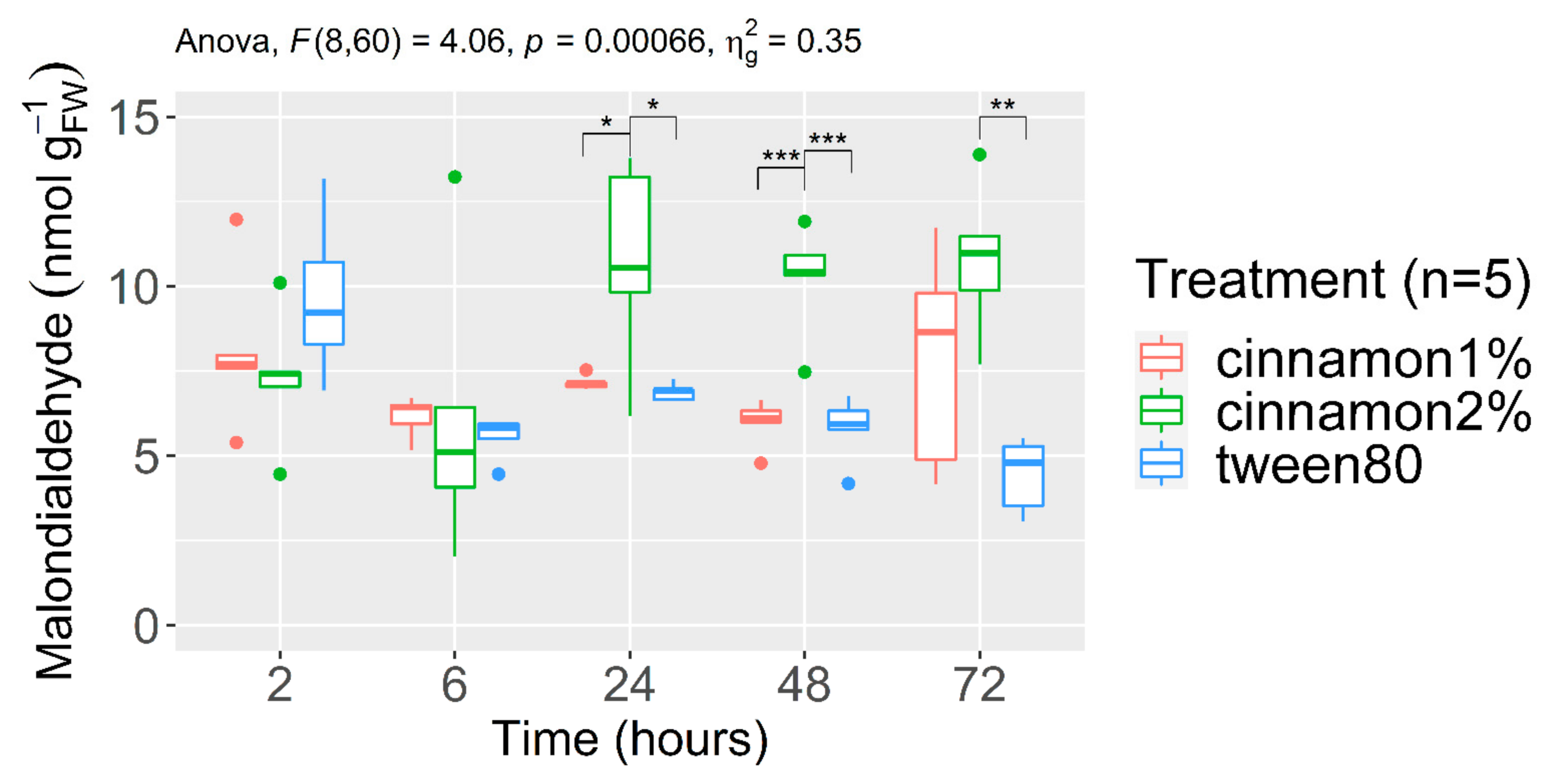
3.2.1. Malondialdehyde Content (MDA)

The bulk of MDA in leaf tissue originates from the poly-unsaturated fatty acids’ (PUFAs) peroxidation in response to oxidative stress. Its content was monitored by the TBARS assay, combined with a final HPLC-DAD separation step. The results following C. cassia EO or tween 80 applications are shown in Figure 2. The main conclusion that can be drawn from these results is that, while the MDA concentration seems to fluctuate between 0 and 6 h of treatment, it increases drastically after 24 h to reach 10.7 ± 3.05 nmol g−1FW for the 2% concentration of CEO. In view of this trend, we can confirm that the peroxidation of membrane lipids causing MDA production would occur between 6 and 24 h after treatment with 2% CEO. For the data that presented a positive skewness, a square-root transformation was applied for statistical analysis. The two-way ANOVA displayed on the top of the graph represents significant interaction between the treatment and time implying that the treatment effect is also time dependent. Results of the pairwise t-test confirmed that from 24 h to 72 h, the 2% CEO treatment modality displays significantly higher values of MDA content compared to the other modalities. This result shows that the antioxidant capacities were not sufficient to inhibit the MDA accumulation in plant. cells.

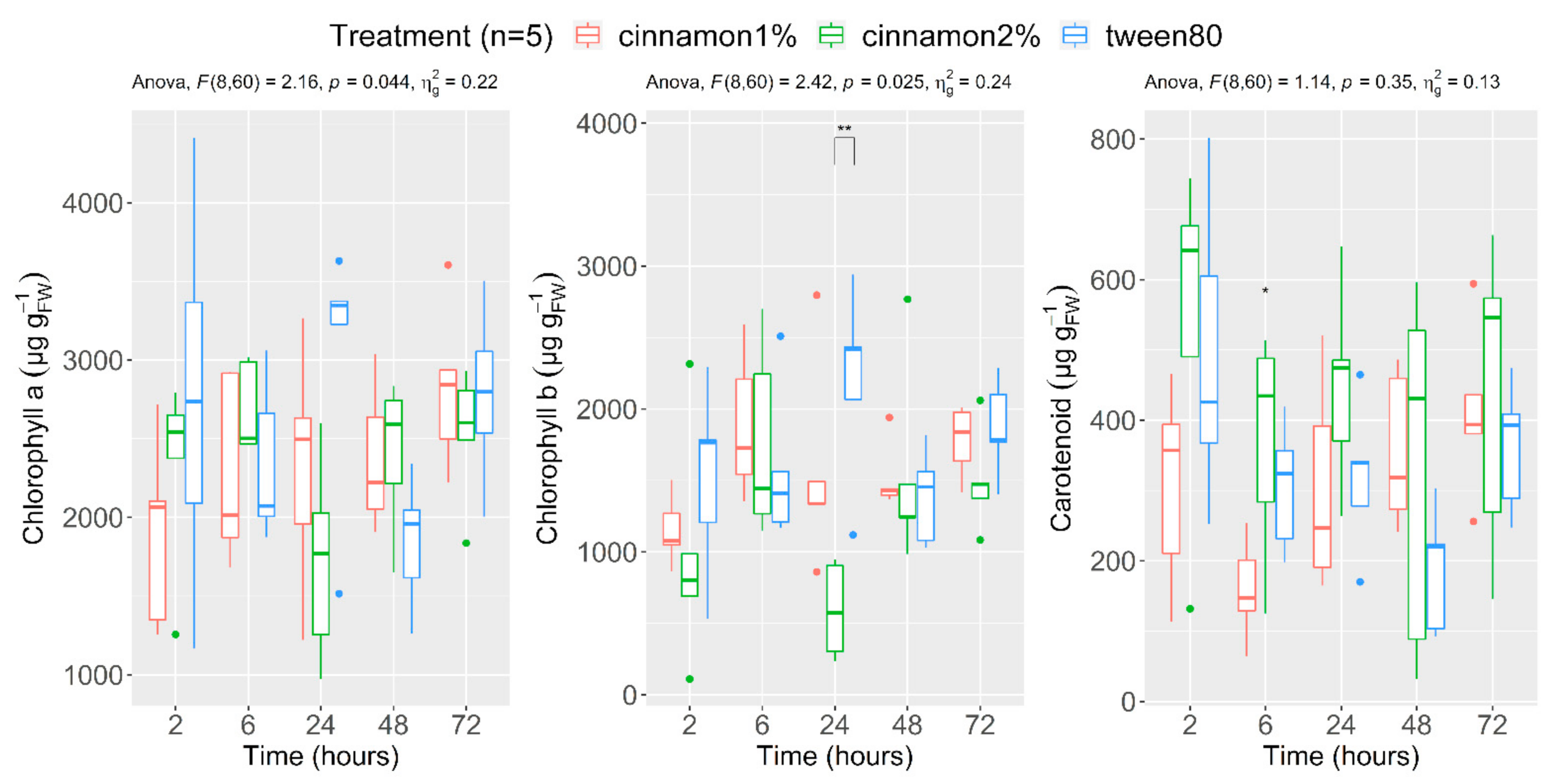
3.2.2. Photosynthetic Pigment Content (Chlorophyll a, Chlorophyll b and Carotenoids)

To follow the potential photosynthetic pigment degradation resulting from CEO application, chlorophyll a, chlorophyll b and carotenoids were measured by spectroscopy in apple leaf ethanolic extract. Their respective contents following CEO application are presented in Figure 3. Chlorophyll a and b contents are in agreement with the literature, with on average, twice the amount of chlorophyll a than b [61]. Moreover, Chl a and b content shows quite a similar tendency, with values sharply decreasing for the 2% concentration after 24 h of treatment and increasing again after 48 h to finally reach initial values. This could be a sign that plant stress management is achieved after 48 h. The two-way ANOVA displayed on the top of the graph demonstrate significant interaction between the treatment and time for Chl a and b. The previous hypothesis is confirmed by statistical analysis only for chlorophyll b, with the 24-h CEO 2% treated plants significantly different from all others. Concerning the carotenoids, the trend is quite different, with values remaining broadly stable from one time step to the next. There is no significant observable difference over time apart for the significantly higher content of the 2% CEO treatment modality after 6 h.

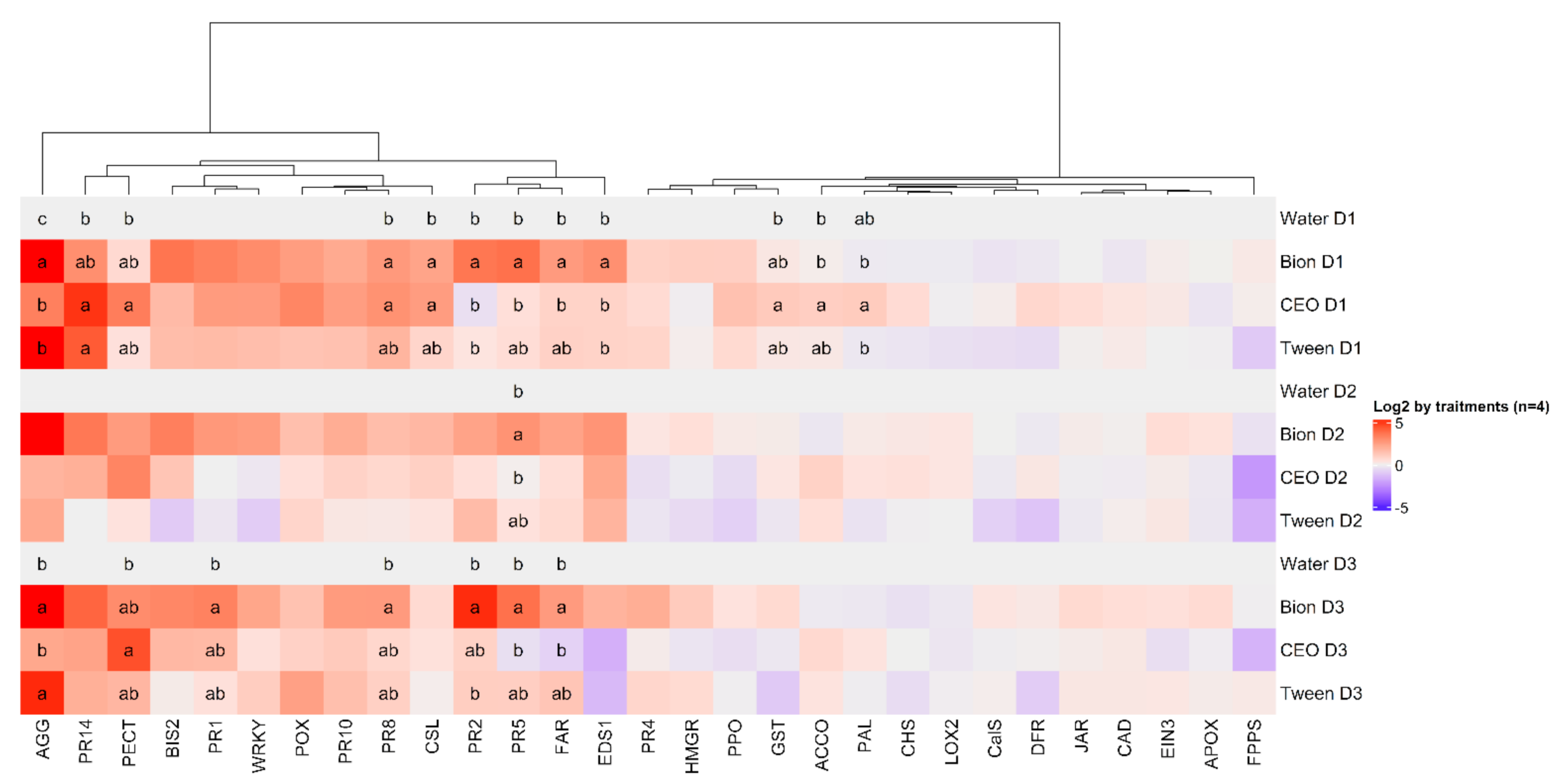
3.3. Induction of Defences

Modification of cellular redox state as well as alteration in the previously mentioned metabolites can lead to reprogramming the expression of diverse genes. To investigate this transcriptional reprogramming, we have applied quantitative real time polymerase chain reaction (qRT-PCR) techniques on 29 transcripts of chemical and physical barriers (PR proteins, phenylpropanoids, isoprenoids, cysteines, oxidative stress, parietal modification and hormonal signalling (salicylic acid (SA), jasmonic acid (JA) and ethylene (ET)). Their detailed codes and names can be found in Table S1. The different treatments consisted of foliar application on apple seedlings (4–6 leaves, from open-pollinated M. domestica cv Golden Delicious) of: Bion® 50 WG (salicylic acid analogue), Tween 80 aqueous solution (surfactant 2%), and emulsions of CEO at 1% (v/v) concentration (the 2% concentration proving to be phytotoxic). The sampling was performed after one, two or three days (corresponding to D1, D2 and D3). Four biological replicates of the same modalities (pooling of five apple seedlings each) were carried out. Principal component analysis (PCA) was performed to investigate the treatments’ impacts on the whole expression profile and representations of daily mean barycenters (with confidence intervals) are displayed in Figure 4 (left). The first two dimensions accounted for 53.5% of the total variability. Initial variable contribution to those dimensions are represented on a variable factor map (right). Regarding barycenters, they separate remarkably following treatment, as illustrated by the confidence intervals. Bion parts following the first dimension whose variable contributions are mostly SAR-related genes such as PR-proteins (PR-1, PR-10, PR-14), oxidative stress (GST, POX), isoprenoids (Far, HMGR) and SA signalling (WRKY EDS1). Tween 80 is closer to the initial time before treatment (T0) and water. Those samples located left (negative value of first dimensions) imply an absence of up-regulation of the previously cited genes. Lastly, the impact of 1% CEO can be highlighted, especially at day 1 (D1). Indeed, it separates close to Bion following the first dimension with up-regulation of SAR-related genes. However, opposite to Bion, this up-regulation of defence genes diminishes drastically after 3 days. Regarding the second axis, no clear features can be underlined. In the PCA, FPPS and EIN3 are the strongest contributors to axis 2 (PC2). These defence genes can also respond strongly to environmental conditions. Therefore, PC2 represents above all a manipulative or sampling day effect, while axis 1 clearly represents the treatment effect.
Figure 5 is a gene expression heatmap representing the mean deviations in the water controls at each sampling date for the 29 defence genes considered, with all values normalised to the initial treatment (represented on Table S2). Among the interesting information that can be derived from this figure, the first is that the control Tween 80 alone produces effects compared to water treatment, that are quite marked for PR proteins and agglutinin. However, Tween 80 has been assessed to be a nontoxic and biocompatible surfactant [62]. Concerning CEO, the activation effects are visible especially at day 1 as evidenced previously in the PCA. Prolonged activation effects until day 3 are observed for some genes, notably for hormonal signalling (ACCO), for pathogenesis-related protein (PR8, PR10 and PR14), for parietal modification (Pect) and phenylpropanoids (BIS2). Bion (Acibenzolar-S-methyl) the positive control, clearly triggers SAR-related genes. Due to their non-normal distribution, impact of treatment was analysed by the Kruskal–Wallis test and post hoc Conover test for pairwise comparison between treatments. We can see significant impacts on each day and between treatments. Indeed, Bion upregulated the following genes (expression level) on day 1, PR-2 (4.24 ± 0.47), PR-5 (3.7 ± 0.68), PR-8 (1.44 ± 0.86), AGG (7.95 ± 1.28), Far (3.47 ± 0.27), CSL (1.72 ± 0.27) and EDS1 (2.61 ± 0.28). On day 2 this increase is significant only for PR-5 (3.19 ± 0.41). Finally, this increase is prolonged until day 3 for PR1 (2.77 ± 0.31), PR2 (4.9 ± 0.33), PR5 (3.48 ± 0.8), PR8 (2.46 ± 0.62), AGG (7.29 ± 1.43) and FAR (2.48 ± 0.54). Tween80 produced a significant increase on day1 for PR-14 (3.44 ± 0.27) and on day 3 for AGG (4.59 ± 0.2). CEO upregulated PR-8 (1.67 ± 0.53), PR-14 (4.14 ± 0.92), PAL (1.56 ± 0.18), CSL (1.95 ± 0.73), GST (0.51 ± 0.17) and ACCO (0.69 ± 0.21) on day 1. Increases are prolonged on day 3 for Pect expression levels (3.43 ± 0.43). Taken individually CEO specifically up-regulated transcripts from different pathway compared to Bion such as ethylene from hormonal signalling (ACCO), oxidative stress (GST) and especially phenylpropanoids (PAL) and parietal modifications (PECT).

4. Discussion

As for many other EOs it seems that foliar application of CEO triggers an oxidative burst as suggested by the drastic decrease in reduced GSH and ratios observed. Whether CEO directly triggers the production of ROS or results from metabolic alteration cannot be deduced from the present study. The pro-oxidative character of CEO depends on its concentration. From a broader perspective, this pro-oxidative character may differ depending on plant sensitivity, tissue type, physiological and/or phenological state. Plants are equipped with numerous soluble antioxidants and many ROS scavenging enzymes (superoxide dismutase (SOD), catalase (CAT), peroxidase (POX), thioredoxin (Trx), and the enzymes of the Asada–Halliwell–Foyer pathway [63]. Their simultaneous measurement gives a better insight of the cellular redox state, but is labour and cost intensive [55]. Glutathione is at the heart of the antioxidant systems. Therefore, GSH redox state measurements have been proposed as a preferred marker for H2O2 availability in plant cells [22], since it has been used to monitor many abiotic stresses, including in apple trees, such as heavy metal [64], drought [65] and temperature [66]. In this framework we propose to include the glutathione redox state measurements as early markers of oxidative burst following EO or VOC applications. Another function of glutathione is to detoxify xenobiotics in plants through adduct formation. These reactions were observed in planta for hexenal [67], methacrolein [68] and are suggested as a conversion method for volatile organic compounds(VOCs) in plant–plant communication [69]. This reaction could also take place and explain part of the GSH consumption.
If not handled by the previously described antioxidant systems, oxidative burst can lead to the appearance of oxidative damage in many macromolecules and in cell membranes, leading to MDA production. MDA Leaf content reported in apple leaf ranges from pmol g−1FW to µmol g−1FW depending on the protocol applied. However, a two-fold increase in content following different types of stress has been acknowledged in heavy metal [64], drought [65] and extreme temperature [66] exposure known to trigger ROS production. MDA originates from PUFA and it is well known that in Arabidopsis thaliana leaves for example, mostly linoleic acid and other tri-unsaturated fatty acids are the source of up to 75% of MDA produced [34]. This specificity makes it a limited marker of oxidative damage. Reactive carbonyl species (RCS) production is considered a ubiquitous reaction to oxidative burst. RCS can inactivate chloroplasts and mitochondrial enzymes accelerating oxidative stress and consuming GSH. Therefore, besides MDA, other compounds should be considered, such as acrolein and 4-hydroxy-2-nonenal [70]. Finally, oxidative damage can occur for other biomolecules beside lipids. Protein inhibition [26], microtubule depolarization [71] or DNA damaging [72] properties have been demonstrated for other VOCs. Those reactions lead to long-term phytotoxicity and should also be considered. Indeed, CEO application can be considered phytotoxic in the short-term only at a 2% concentration, but we can’t rule out other mechanisms that lead to long-term phytotoxicity at lower concentrations.
Pathogenesis-Related (PR) proteins have been defined as plant host proteins that are produced only in response to attack by pathogens or a related event [73]. Demonstrating the expression of PR genes has been widely accepted as a hallmark of plant defensive systemic acquired resistance (SAR) induction [74,75]. The SAR, which is a form of systemic resistance in plants with a specific defence signalling pathway, can also occur after spraying with a synthetic or natural compound, commonly known as an inducer such as the Bion used in this study [47]. From our results (PCA) it would seem that defence induction pathways following CEO application is similar to SAR. The most commonly screened PR genes expressed in apples and other plant-pathogen systems are PR-1 (antifungal activity), PR-2 (β-1,3-glucanase), and PR-8 (class III chitinase) [76]. Thyme EO has been suggested to increase PR-8 expression in apple [39]. Our results also showed significant increases in expression levels of PR-8 and PR-14 compared to water. Tween 80 alone also impacted PR-related protein agglutinin and PR-14. Similar results on defence related genes have been previously highlighted in wheat after tween 20 treatment [77]. Therefore, the formulation needs to be investigated to determine if defence induction is actually due to the elicitor compounds themselves.
Apart from those coding for PR proteins, other genes represent the wide diversity of known plant defence mechanisms. The metabolic pathways to which these genes are related include secondary metabolic pathways (phenylpropanoids and isoprenoids), oxidative stress, parietal modifications and hormonal signalling pathways of salicylic acid, jasmonic acid and ethylene. In our study, phenylalanine ammonia-lyase (PAL) expression levels from the phenylpropanoids pathway was significantly (but transiently) increased. This is coherent with previous results obtained regarding this enzyme activity in citrus following trans-cinnamaldehyde application [45]. Changes in PAL activity have been shown to precede the increases in BD and BIS activities in Asian pear Pyrus pyrifolia leading to production of phytoalexins [78]. This result was not verified here, but only BIS2 was followed out of the 9 genes detected in the genome sequence of the apple ‘Golden Delicious’ [79]. Alternatively, phenylpropanoid pathway activation could be investigated through production of biphenyls and dibenzofurans. Indeed, production of aucuparin and noraucuparin have been demonstrated in apple following elicitor treatment [80]. Cell wall modification may also occur, as suggested by the increase in pectin methyl esterase (PECT) expression levels. Regarding hormonal signalling, ACCO up-regulation suggests an impact on ethylene. Hormonal signalling is known to be a very transient signal; therefore, the balance of phytohormones should be considered when investigating signal perception following CEO application. Concerning oxidative stress, glutathione S-transferase (GST) is specifically up-regulated after CEO application which is consistent with the GSH results presented, regarding redox status. Finally, regarding isoprenoids, α-farnesene production was acknowledged in response to SAR induction by Bion application. The same result was obtained following CEO injection into apple trees with a modification of VOC emission [81]. However Farnesyl pyrophosphate synthase (FPPS) and (E,E)-α-farnesene synthase (FAR) do not seem to be upregulated following CEO application. However, deep transcriptome analyses such as RNA-seq should provide a more complete and less biased picture on all the genes modulated following CEO treatment.
Regarding signal transduction, it has been suggested that while the monoterpenes could disturb the lipid organization and/or domain formation, the phenylpropanoid cinnamaldehyde could rather interact with membrane receptors [49]. Recent evidence suggests that cinnamaldehyde regulates endogenous Ca2+ in the root of Brassica rapa [82]. Furthermore, investigation observed generation of endogenous hydrogen sulphide (H2S) in roots treated with cinnamaldehyde, supposedly by increasing the activity of L-cysteine desulfhydrase [83]. They proposed that cinnamaldehyde could regulate Ca2+ directly by targeting transient receptor potential A1 (TRPA1) as observed in mammals, but also that Ca2+ regulation by H2S may operate downstream of cinnamaldehyde through a linear signalling pathway during the induction of lateral root formation. The transcriptional reprogramming that follows CEO application could be explained by modification of those prominent signalling molecules.
To conclude, this work highlights modification of oxidative stress related metabolites; namely glutathione and malondialdehyde, following CEO application in a dose–response relationship. Furthermore, it investigates transcriptional reprogramming of the major defence pathway. Increases in expression levels of specific genes belonging to PR-proteins (PR-8, PR-14), hormonal signalling (ACCO), oxidative stress (GST), phenylpropanoids (PAL) and parietal modification (PECT) pathways were observed following CEO application. Multivariate analysis of the 29 transcripts acknowledged similar but more transient modification of expression levels than the SAR inducer Bion.
In a broader scope, the defence induction occurring below the phytotoxicity threshold represents an engaging research path for EO application in agronomy to design appropriate and sustainable agricultural pest management strategies.

Supplementary Materials

The following are available online at https://www.mdpi.com/article/10.3390/agronomy12020512/s1. Figure S1: Typical HPLC–FLD sample chromatogram (RT GSH–MBB = 7.2min) and calibration curve for GSH; Limit of detection (LOD) = 1.16 µM; Limit of quantification (LOQ) = 1.41 µM; Figure S2: Typical HPLC–DAD sample chromatogram (RT MDA(TBA)2 = 2.1min) chromatogram and calibration curve for MDA; Limit of detection (LOD) = 0.19 µM; Limit of quantification (LOQ) = 0.25 µM; Table S1: List of 29 defence genes followed by qRT-PCR; Table S2: Log 2 expression level of 29 defence genes followed by qRT-PCR.

Author Contributions

Conceptualization, P.-Y.W. and M.-L.F.; methodology, P.-Y.W.; software, P.-Y.W.; validation, P.-Y.W., M.-L.F. and M.-N.B.; formal analysis, P.-Y.W., C.H. and A.J.; data curation, P.-Y.W. and A.J.; writing—original draft preparation, P.-Y.W.; writing—review and editing, P.-Y.W., A.J., M.-L.F. and M.-N.B.; supervision, M.-L.F.; project administration, M.-L.F.; funding acquisition, M.-L.F. All authors have read and agreed to the published version of the manuscript.

Funding

This research was funded by the Department of Research and Technological Development of the Walloon regions of Belgium (DG03) through the TREE-INJECTION project R. RWAL-3157.

Institutional Review Board Statement

Not applicable.

Informed Consent Statement

Not applicable.

Data Availability Statement

The data are available on request from the corresponding author.

Acknowledgments

We thank Danny Trisman and Thomas Bertrand for their technical contribution, Laura Lheureux and Jean Bouvry for their contribution by their master thesis.

Conflicts of Interest

The authors declare no conflict of interest.

References

  1. O’Bryan, C.A.; Pendleton, S.J.; Crandall, P.G.; Ricke, S.C. Potential of plant essential oils and their components in animal agriculture-in vitro studies on antibacterial mode of action. Front. Vet. Sci. 2015, 2, 35. [Google Scholar] [CrossRef] [PubMed] [Green Version]
  2. De Clerck, C.; Maso, S.D.; Parisi, O.; Dresen, F.; Zhiri, A.; Haissam Jijakli, M. Screening of antifungal and antibacterial activity of 90 commercial essential oils against 10 pathogens of agronomical importance. Foods 2020, 9, 1418. [Google Scholar] [CrossRef] [PubMed]
  3. Jankowska, M.; Rogalska, J.; Wyszkowska, J.; Stankiewicz, M. Molecular targets for components of essential oils in the insect nervous system—A review. Molecules 2018, 23, 34. [Google Scholar] [CrossRef] [PubMed] [Green Version]
  4. Peixoto, M.G.; Costa-Júnior, L.M.; Blank, A.F.; Lima, A.d.S.; Menezes, T.S.A.; Santos, D.d.A.; Alves, P.B.; Cavalcanti, S.C.d.H.; Bacci, L.; Arrigoni-Blank, M.d.F. Acaricidal activity of essential oils from Lippia alba genotypes and its major components carvone, limonene, and citral against Rhipicephalus microplus. Vet. Parasitol. 2015, 210, 118–122. [Google Scholar] [CrossRef] [PubMed]
  5. Andrés, M.F.; González-Coloma, A.; Sanz, J.; Burillo, J.; Sainz, P. Nematicidal activity of essential oils: A review. Phytochem. Rev. 2012, 11, 371–390. [Google Scholar] [CrossRef] [Green Version]
  6. Raveau, R.; Fontaine, J.; Lounès-Hadj Sahraoui, A. Essential oils as potential alternative biocontrol products against plant pathogens and weeds: A review. Foods 2020, 9, 365. [Google Scholar] [CrossRef] [Green Version]
  7. Dayan, F.E.; Cantrell, C.L.; Duke, S.O. Natural products in crop protection. Bioorg. Med. Chem. 2009, 17, 4022–4034. [Google Scholar] [CrossRef]
  8. Coats, J.R.; Karr, L.L.; Drewes, C.D. Toxicity and Neurotoxic Effects of Monoterpenoids: In Insects and Earthworms. Nat. Occur. Pest Bioregulators 1991, 449, 305. [Google Scholar]
  9. Kostyukovsky, M.; Rafaeli, A.; Gileadi, C.; Demchenko, N.; Shaaya, E. Activation of octopaminergic receptors by essential oil constituents isolated from aromatic plants: Possible mode of action against insect pests. Pest Manag. Sci. 2002, 58, 1101–1106. [Google Scholar] [CrossRef] [PubMed]
  10. Mossa, A.T.H. Green Pesticides: Essential oils as biopesticides in insect-pest management. J. Environ. Sci. Technol. 2016, 9, 354–378. [Google Scholar] [CrossRef] [Green Version]
  11. Pavela, R.; Benelli, G. Essential Oils as Ecofriendly Biopesticides? Challenges and Constraints. Trends Plant Sci. 2016, 21, 1000–1007. [Google Scholar] [CrossRef] [PubMed]
  12. Renoz, F.; Demeter, S.; Degand, H.; Nicolis, S.C.; Lebbe, O.; Martin, H.; Deneubourg, J.-L.; Fauconnier, M.L.; Morsomme, P.; Hance, T. The modes of action of Mentha arvensis essential oil on the granary weevil Sitophilus granarius revealed by a label-free quantitative proteomic analysis. J. Pest Sci. 2022, 95, 381–395. [Google Scholar] [CrossRef]
  13. Werrie, P.-Y.; Durenne, B.; Delaplace, P.; Fauconnier, M.-L. Phytotoxicity of Essential Oils: Opportunities and Constraints for the Development of Biopesticides. A Review. Foods 2020, 9, 1291. [Google Scholar] [CrossRef] [PubMed]
  14. Mittler, R.; Vanderauwera, S.; Gollery, M.; Van Breusegem, F. Reactive oxygen gene network of plants. Trends Plant Sci. 2004, 9, 490–498. [Google Scholar] [CrossRef] [PubMed]
  15. Gill, S.S.; Anjum, N.A.; Hasanuzzaman, M.; Gill, R.; Trivedi, D.K.; Ahmad, I.; Pereira, E.; Tuteja, N. Glutathione and glutathione reductase: A boon in disguise for plant abiotic stress defense operations. Plant Physiol. Biochem. 2013, 70, 204–212. [Google Scholar] [CrossRef] [PubMed]
  16. Noctor, G.; Queval, G.; Mhamdi, A.; Chaouch, S.; Foyer, C.H. Glutathione. Arab. B. 2011, 9, e0142. [Google Scholar] [CrossRef]
  17. Queval, G.; Jaillard, D.; Zechmann, B.; Noctor, G. Increased intracellular H2O2 availability preferentially drives glutathione accumulation in vacuoles and chloroplasts. Wiley Online Libr. 2010, 34, 21–32. [Google Scholar] [CrossRef]
  18. Hajdinák, P.; Czobor, Á.; Lőrincz, T.; Szarka, A. The problem of glutathione determination: A comparative study on the measurement of glutathione from plant cells. Period. Polytech. Chem. Eng. 2019, 63, 1–10. [Google Scholar] [CrossRef]
  19. Davey, M.W.; Dekempeneer, E.; Keulemans, J. Rocket-powered high-performance liquid chromatographic analysis of plant ascorbate and glutathione. Anal. Biochem. 2003, 316, 74–81. [Google Scholar] [CrossRef]
  20. Noctor, G.; Mhamdi, A.; Chaouch, S.; Han, Y.; Neukermans, J.; Marquez-Garcia, B.; Queval, G.; Foyer, C.H. Glutathione in plants: An integrated overview. Plant Cell Environ. 2012, 35, 454–484. [Google Scholar] [CrossRef]
  21. Gniazdowska, A.; Krasuska, U.; Andrzejczak, O.; Soltys, D. Allelopathic Compounds as Oxidative Stress Agents: Yes or NO. In Signaling and Communication in Plants; Springer: Cham, Switzerland, 2015; Volume 23, pp. 155–176. [Google Scholar]
  22. Foyer, C.H.; Lopez-Delgado, H.; Dat, J.F.; Scott, I.M. Hydrogen peroxide- and glutathione-associated mechanisms of acclimatory stress tolerance and signalling. Physiol. Plant. 1997, 100, 241–254. [Google Scholar] [CrossRef]
  23. Morales, M.; Munné-Bosch, S. Malondialdehyde: Facts and artifacts. Plant Physiol. 2019, 180, 1246–1250. [Google Scholar] [CrossRef] [PubMed] [Green Version]
  24. Davey, M.W.; Stals, E.; Panis, B.; Keulemans, J.; Swennen, R.L. High-throughput determination of malondialdehyde in plant tissues. Anal. Biochem. 2005, 347, 201–207. [Google Scholar] [CrossRef] [PubMed]
  25. Miguel, M.G. Antioxidant and anti-inflammatory activities of essential oils: A short review. Molecules 2010, 15, 9252–9287. [Google Scholar] [CrossRef] [PubMed] [Green Version]
  26. Araniti, F.; Landi, M.; Lupini, A.; Sunseri, F.; Guidi, L.; Abenavoli, M.R. Origanum vulgare essential oils inhibit glutamate and aspartate metabolism altering the photorespiratory pathway in Arabidopsis thaliana seedlings. J. Plant Physiol. 2018, 231, 297–309. [Google Scholar] [CrossRef] [PubMed]
  27. Pouresmaeil, M.; Nojadeh, M.S.; Movafeghi, A.; Maggi, F. Exploring the bio-control efficacy of Artemisia fragrans essential oil on the perennial weed Convolvulus arvensis: Inhibitory effects on the photosynthetic machinery and induction of oxidative stress. Ind. Crops Prod. 2020, 155, 112785. [Google Scholar] [CrossRef]
  28. Poonpaiboonpipat, T.; Pangnakorn, U.; Suvunnamek, U.; Teerarak, M.; Charoenying, P.; Laosinwattana, C. Phytotoxic effects of essential oil from Cymbopogon citratus and its physiological mechanisms on barnyardgrass (Echinochloa crus-galli). Ind. Crops Prod. 2013, 41, 403–407. [Google Scholar] [CrossRef]
  29. Gao, Y.; Kan, C.; Chen, M.; Chen, C.; Chen, Y.; Fu, Y.; Wan, C.; Chen, J. Effects of Chitosan-Based Coatings Enriched with Cinnamaldehyde on Mandarin Fruit cv. Ponkan during Room-Temperature Storage. Coatings 2018, 8, 372. [Google Scholar] [CrossRef]
  30. Chowhan, N.; Singh, H.P.; Batish, D.R.; Kohli, R.K. Phytotoxic effects of β-pinene on early growth and associated biochemical changes in rice. Acta Physiol. Plant. 2011, 33, 2369–2376. [Google Scholar] [CrossRef]
  31. Bresson, J.; Bieker, S.; Riester, L.; Doll, J.; Zentgraf, U. A guideline for leaf senescence analyses: From quantification to physiological and molecular investigations. J. Exp. Bot. 2018, 69, 769–786. [Google Scholar] [CrossRef]
  32. Radhakrishnan, R.; Alqarawi, A.A.; Abd Allah, E.F. Bioherbicides: Current knowledge on weed control mechanism. Ecotoxicol. Environ. Saf. 2018, 158, 131–138. [Google Scholar] [CrossRef] [PubMed]
  33. Gitelson, A.A.; Merzlyak, M.N. Non-destructive assessment of chlorophyll, carotenoid and anthocyanin in higher plant leaves: Principles and algorithms. Remote Sens. Agric. Environ. 2004, 78–94. Available online: https://digitalcommons.unl.edu/cgi/viewcontent.cgi?article=1265&context=natrespapers (accessed on 10 January 2022).
  34. Weber, H.; Chételat, A.; Reymond, P.; Farmer, E.E. Selective and powerful stress gene expression in Arabidopsis in response to malondialdehyde. Plant J. 2004, 37, 877–888. [Google Scholar] [CrossRef] [PubMed]
  35. Han, Y.; Mhamdi, A.; Chaouch, S.; Noctor, G. Regulation of basal and oxidative stress-triggered jasmonic acid-related gene expression by glutathione. Plant Cell Environ. 2013, 36, 1135–1146. [Google Scholar] [CrossRef]
  36. Gomez, L.D.; Noctor, G.; Knight, M.R.; Foyer, C.H. Regulation of calcium signalling and gene expression by glutathione. J. Exp. Bot. 2004, 55, 1851–1859. [Google Scholar] [CrossRef] [Green Version]
  37. Massimo, E. Maffei Monoterpenoid Plant-Plant Interactions Upon Herbivory. Curr. Bioact. Compd. 2012, 8, 65–70. [Google Scholar] [CrossRef]
  38. Ben-Jabeur, M.; Kthiri, Z.; Harbaoui, K.; Belguesmi, K.; Serret, M.D.; Araus, J.L.; Hamada, W. Seed Coating with Thyme Essential Oil or Paraburkholderia phytofirmans PsJN Strain: Conferring Septoria Leaf Blotch Resistance and Promotion of Yield and Grain Isotopic Composition in Wheat. Agronomy 2019, 9, 586. [Google Scholar] [CrossRef] [Green Version]
  39. Banani, H.; Olivieri, L.; Santoro, K.; Garibaldi, A.; Gullino, M.; Spadaro, D. Thyme and Savory Essential Oil Efficacy and Induction of Resistance against Botrytis cinerea through Priming of Defense Responses in Apple. Foods 2018, 7, 11. [Google Scholar] [CrossRef] [Green Version]
  40. Sukegawa, S.; Shiojiri, K.; Higami, T.; Suzuki, S.; Arimura, G.-i. Pest management using mint volatiles to elicit resistance in soy: Mechanism and application potential. Plant J. 2018, 96, 910–920. [Google Scholar] [CrossRef] [Green Version]
  41. Godard, K.A.; White, R.; Bohlmann, J. Monoterpene-induced molecular responses in Arabidopsis thaliana. Phytochemistry 2008, 69, 1838–1849. [Google Scholar] [CrossRef]
  42. Chen, S.; Zhang, L.; Cai, X.; Li, X.; Bian, L.; Luo, Z.; Li, Z.; Chen, Z.; Xin, Z. (E)-Nerolidol is a volatile signal that induces defenses against insects and pathogens in tea plants. Hortic. Res. 2020, 7, 52. [Google Scholar] [CrossRef] [PubMed] [Green Version]
  43. Ben-Jabeur, M.; Ghabri, E.; Myriam, M.; Hamada, W. Thyme essential oil as a defense inducer of tomato against gray mold and Fusarium wilt. Plant Physiol. Biochem. 2015, 94, 35–40. [Google Scholar] [CrossRef]
  44. Jhonata, L.d.S.; Paulo, E.d.S.; Eduardo, A.; Jose, E.B.P.P.; Suzan, K.V.B.; Marcelo, L.O.F.; Camila, C.L.d.A.; Mario, L.V.R. Essential oil of Cymbopogon flexuosus, Vernonia polyanthes and potassium phosphite in control of bean anthracnose. J. Med. Plants Res. 2015, 9, 243–253. [Google Scholar] [CrossRef]
  45. Perina, F.J.; de Andrade, C.C.L.; Moreira, S.I.; Nery, E.M.; Ogoshi, C.; Alves, E. Cinnamomun zeylanicum oil and trans-cinnamaldehyde against Alternaria brown spot in tangerine: Direct effects and induced resistance. Phytoparasitica 2019, 47, 575–589. [Google Scholar] [CrossRef]
  46. Pereira, R.B.; Lucas, G.C.; Perina, F.J.; Ribeiro, P.M., Jr.; Alves, E. Citronella essential oil in the control and activation of coffee plants defense response against rust and brown eye spot. Ciência Agrotecnol. 2012, 36, 383–390. [Google Scholar] [CrossRef] [Green Version]
  47. Dugé De Bernonville, T.; Marolleau, B.; Staub, J.; Gaucher, M.; Brisset, M.N. Using molecular tools to decipher the complex world of plant resistance inducers: An apple case study. J. Agric. Food Chem. 2014, 62, 11403–11411. [Google Scholar] [CrossRef]
  48. Koul, O.; Walia, S.; Dhaliwal, G. Essential oils as green pesticides: Potential and Constraints. Biopestic. Int. 2008, 4, 63–84. [Google Scholar]
  49. Lins, L.; Dal Maso, S.; Foncoux, B.; Kamili, A.; Laurin, Y.; Genva, M.; Jijakli, M.H.; De Clerck, C.; Fauconnier, M.L.; Deleu, M. Insights into the Relationships Between Herbicide Activities, Molecular Structure and Membrane Interaction of Cinnamon and Citronella Essential Oils Components. Int. J. Mol. Sci. 2019, 20, 4007. [Google Scholar] [CrossRef] [Green Version]
  50. Mezőfi, L.; Sipos, P.; Vétek, G.; Elek, R.; Markó, V. Evaluation of kaolin and cinnamon essential oil to manage two pests and a fungal disease of sour cherry at different tree canopy levels. J. Plant Dis. Prot. 2018, 125, 483–490. [Google Scholar] [CrossRef]
  51. Washington, D.C.; Mavian, C.M.K. U.S. EPA, Pesticide Product Label, CINNACURE P1005, 01/21/1999. 1999. Available online: https://www3.epa.gov/pesticides/chem_search/ppls/058866-00011-19990121.pdf (accessed on 2 February 2022).
  52. Warneys, R.; Gaucher, M.; Robert, P.; Aligon, S.; Anton, S.; Aubourg, S.; Barthes, N.; Braud, F.; Cournol, R.; Gadenne, C.; et al. Acibenzolar-s-methyl reprograms apple transcriptome toward resistance to rosy apple aphid. Front. Plant Sci. 2018, 871, 1795. [Google Scholar] [CrossRef]
  53. Le Mire, G.; Siah, A.; Marolleau, B.; Gaucher, M.; Maumené, C.; Brostaux, Y.; Massart, S.; Brisset, M.N.; Haissam Jijakli, M. Evaluation of l-carrageenan, CpG-ODN, glycine betaine, spirulina platensis, and ergosterol as elicitors for control of zymoseptoria tritici in wheat. Phytopathology 2019, 109, 409–417. [Google Scholar] [CrossRef] [PubMed] [Green Version]
  54. Le Mire, G.; Siah, A.; Brisset, M.N.; Gaucher, M.; Deleu, M.; Jijakli, M.H. Surfactin protects wheat against Zymoseptoria tritici and activates both salicylic acid- and jasmonic acid-dependent defense responses. Agriculture 2018, 8, 11. [Google Scholar] [CrossRef] [Green Version]
  55. Queval, G.; Noctor, G. A plate reader method for the measurement of NAD, NADP, glutathione, and ascorbate in tissue extracts: Application to redox profiling during Arabidopsis rosette development. Anal. Biochem. 2007, 363, 58–69. [Google Scholar] [CrossRef] [PubMed]
  56. Velikova, V.; Yordanov, I.; Edreva, A. Oxidative stress and some antioxidant systems in acid rain-treated bean plants protective role of exogenous polyamines. Plant Sci. 2000, 151, 59–66. [Google Scholar] [CrossRef]
  57. Vergnes, S.; Ladouce, N.; Fournier, S.; Ferhout, H.; Attia, F.; Dumas, B. Foliar treatments with Gaultheria procumbens essential oil induce defense responses and resistance against a fungal pathogen in Arabidopsis. Front. Plant Sci. 2014, 5, 477. [Google Scholar] [CrossRef]
  58. Bernonville, T.D.d.; Gaucher, M.; Guyot, S.; Durel, C.E.; Dat, J.F.; Brisset, M.N. The constitutive phenolic composition of two Malus×domestica genotypes is not responsible for their contrasted susceptibilities to fire blight. Environ. Exp. Bot. 2011, 74, 65–73. [Google Scholar] [CrossRef] [Green Version]
  59. Gallé, Á.; Czékus, Z.; Bela, K.; Horváth, E.; Ördög, A.; Csiszár, J.; Poór, P. Plant glutathione transferases and light. Front. Plant Sci. 2019, 9, 1944. [Google Scholar] [CrossRef] [Green Version]
  60. Meyer, A.J. The integration of glutathione homeostasis and redox signaling. J. Plant Physiol. 2008, 165, 1390–1403. [Google Scholar] [CrossRef]
  61. Tamburini, E.; Ferrari, G.; Marchetti, M.; Pedrini, P.; Ferro, S. Development of FT-NIR models for the simultaneous estimation of chlorophyll and nitrogen content in fresh apple (Malus domestica) leaves. Sensors 2015, 15, 2662–2679. [Google Scholar] [CrossRef] [Green Version]
  62. Prieto, C.; Calvo, L. Performance of the Biocompatible Surfactant Tween 80, for the Formation of Microemulsions Suitable for New Pharmaceutical Processing. J. Appl. Chem. 2013, 2013, 930356. [Google Scholar] [CrossRef] [Green Version]
  63. Foyer, C.H.; Noctor, G. Redox Homeostasis and Antioxidant Signaling: A Metabolic Interface between Stress Perception and Physiological Responses. Plant Cell 2005, 17, 1866–1875. [Google Scholar] [CrossRef] [PubMed] [Green Version]
  64. He, J.; Zhuang, X.; Zhou, J.; Sun, L.; Wan, H.; Li, H.; Lyu, D. Exogenous melatonin alleviates cadmium uptake and toxicity in apple rootstocks. Tree Physiol. 2020, 40, 746–761. [Google Scholar] [CrossRef] [PubMed]
  65. Ma, Y.H.; Ma, F.W.; Wang, Y.H.; Zhang, J.K. The responses of the enzymes related with ascorbate-glutathione cycle during drought stress in apple leaves. Acta Physiol. Plant. 2011, 33, 173–180. [Google Scholar] [CrossRef]
  66. Ma, Y.H.; Ma, F.W.; Zhang, J.K.; Li, M.J.; Wang, Y.H.; Liang, D. Effects of high temperature on activities and gene expression of enzymes involved in ascorbate–glutathione cycle in apple leaves. Plant Sci. 2008, 175, 761–766. [Google Scholar] [CrossRef]
  67. Davoine, C.; Falletti, O.; Douki, T.; Iacazio, G.; Ennar, N.; Montillet, J.L.; Triantaphylidès, C. Adducts of Oxylipin Electrophiles to Glutathione Reflect a 13 Specificity of the Downstream Lipoxygenase Pathway in the Tobacco Hypersensitive Response. Plant Physiol. 2006, 140, 1484–1493. [Google Scholar] [CrossRef] [Green Version]
  68. Muramoto, S.; Matsubara, Y.; Mwenda, C.M.; Koeduka, T.; Sakami, T.; Tani, A.; Matsui, K. Glutathionylation and reduction of methacrolein in tomato plants account for its absorption from the vapor phase. Plant Physiol. 2015, 169, 1744–1754. [Google Scholar] [CrossRef] [Green Version]
  69. Sugimoto, K.; Matsui, K.; Takabayashi, J. Uptake and Conversion of Volatile Compounds in Plant–Plant Communication. In Deciphering Chemical Language of Plant Communication; Springer: Cham, Switzerland, 2016; pp. 305–316. [Google Scholar]
  70. Mano, J. Reactive carbonyl species: Their production from lipid peroxides, action in environmental stress, and the detoxification mechanism. Plant Physiol. Biochem. 2012, 59, 90–97. [Google Scholar] [CrossRef]
  71. Chaimovitsh, D.; Shachter, A.; Abu-Abied, M.; Rubin, B.; Sadot, E.; Dudai, N. Herbicidal Activity of Monoterpenes Is Associated with Disruption of Microtubule Functionality and Membrane Integrity. Weed Sci. 2017, 65, 19–30. [Google Scholar] [CrossRef] [Green Version]
  72. Bozari, S.; Agar, G.; Aksakal, O.; Erturk, F.A.; Yanmis, D. Determination of chemical composition and genotoxic effects of essential oil obtained from Nepeta nuda on Zea mays seedlings. Toxicol. Ind. Health 2013, 29, 339–348. [Google Scholar] [CrossRef]
  73. Van Loon, L.C.; Pierpoint, W.S.; Boller, T.; Conejero, V. Recommendations for naming plant pathogenesis-related proteins. Plant Mol. Biol. Report. 1994, 12, 245–264. [Google Scholar] [CrossRef]
  74. Bonasera, J.M.; Kim, J.F.; Beer, S.V. PR genes of apple: Identification and expression in response to elicitors and inoculation with Erwinia amylovora. BMC Plant Biol. 2006, 6, 23. [Google Scholar] [CrossRef] [Green Version]
  75. Oliveira, M.D.M.; Varanda, C.M.R.; Félix, M.R.F. Induced resistance during the interaction pathogen x plant and the use of resistance inducers. Phytochem. Lett. 2016, 15, 152–158. [Google Scholar] [CrossRef] [Green Version]
  76. Saboki, E.; Usha, K.; Singh, B. Pathogenesis Related (PR) Proteins in Plant Defense Mechanism Age-Related Pathogen Resistance. Curr. Res. Technol. Adv. 2011, 2, 1043–1054. [Google Scholar]
  77. Le Mire, G. Identification of Elicitors Inducing Resistance in Wheat against Zymoseptoria Tritici and Characterization of the Subsequent Triggered Defense-Signaling Pathways. 2018. Available online: https://orbi.uliege.be/handle/2268/222563 (accessed on 2 February 2022).
  78. Saini, S.S.; Teotia, D.; Gaid, M.; Thakur, A.; Beerhues, L.; Sircar, D. Benzaldehyde dehydrogenase-driven phytoalexin biosynthesis in elicitor-treated Pyrus pyrifolia cell cultures. J. Plant Physiol. 2017, 215, 154–162. [Google Scholar] [CrossRef] [PubMed]
  79. Chizzali, C.; Gaid, M.M.; Belkheir, A.K.; Hänsch, R.; Richter, K.; Flachowsky, H.; Peil, A.; Hanke, M.V.; Liu, B.; Beerhues, L. Differential Expression of Biphenyl Synthase Gene Family Members in Fire-Blight-Infected Apple ‘Holsteiner Cox’. Plant Physiol. 2012, 158, 864–875. [Google Scholar] [CrossRef] [PubMed] [Green Version]
  80. Sarkate, A.; Saini, S.S.; Teotia, D.; Gaid, M.; Mir, J.I.; Roy, P.; Agrawal, P.K.; Sircar, D. Comparative metabolomics of scab-resistant and susceptible apple cell cultures in response to scab fungus elicitor treatment. Sci. Rep. 2018, 8, 17844. [Google Scholar] [CrossRef] [PubMed] [Green Version]
  81. Werrie, P.-Y.; Burgeon, C.; Le Goff, G.J.; Hance, T.; Fauconnier, M.-L. Biopesticide Trunk Injection Into Apple Trees: A Proof of Concept for the Systemic Movement of Mint and Cinnamon Essential Oils. Front. Plant Sci. 2021, 12, 495. [Google Scholar] [CrossRef]
  82. Cheng, Y.; Wang, N.; Liu, R.; Bai, H.; Tao, W.; Chen, J.; Shi, Z. Cinnamaldehyde Facilitates Cadmium Tolerance by Modulating Ca2+ in Brassica rapa. Water. Air. Soil Pollut. 2021, 232, 19. [Google Scholar] [CrossRef]
  83. Xue, Y.F.; Zhang, M.; Qi, Z.Q.; Li, Y.Q.; Shi, Z.; Chen, J. Cinnamaldehyde promotes root branching by regulating endogenous hydrogen sulfide. J. Sci. Food Agric. 2016, 96, 909–914. [Google Scholar] [CrossRef]
Figure 1. Effect of C. cassia EO (CEO) (1 and 2% v/v) and tween 80 application on glutathione leaf content over time (n = 5): (left) Reduced glutathione GSH (nmol g−1FW); (center) Total glutathione GSH+GSSG (nmol g−1FW;) (right) the glutathione ratio GSH/GSH+GSSG (%). Star on boxplot indicates significantly different distributions (* p < 0.05, ** p < 0.01, *** p < 0.001, pairwise t-test).
Agronomy 12 00512 g001
Figure 2. Effect of C. cassia EO (CEO) (1 and 2% v/v) and tween 80 application on malondialdehyde (MDA) leaf content (ng/g) over time (n = 5). Star on boxplot indicates significantly different distributions (* p < 0.05, ** p < 0.01, *** p < 0.001, pairwise t-test).
Agronomy 12 00512 g002
Figure 3. Effect of C. cassia EO (CEO) (1 and 2% v/v) and tween 80 application on photosynthetic pigment leaf content over time (n = 5): (left) Chlorophyll a content (µg/g); (center) Chlorophyll b content (µg/g); (right) Carotenoid content (µg/g). Star on boxplot indicates significantly different distributions (* p < 0.05, ** p < 0.01).
Agronomy 12 00512 g003
Figure 4. Effect of C. cassia EO (CEO 1% (v/v)), Bion, tween 80 and water application on mean normalised log 2 expression level (n = 4) by days of 29 mRNA transcripts of selected defence genes analysed by principal component analysis (PCA) with barycenter representation (left) and variable contribution to dimensions (right).
Agronomy 12 00512 g004
Figure 5. Effect of C. cassia EO (CEO 1% (v/v)), Bion, tween 80 and water application on mean normalised log 2 expression levels (n = 4) by days of 29 mRNA transcripts of selected defence genes analysed by heatmap (deviations to the water controls) and the pairwise Conover test with compact letter displays (different letter representing significantly different mean).
Agronomy 12 00512 g005
Publisher’s Note: MDPI stays neutral with regard to jurisdictional claims in published maps and institutional affiliations.

