Study may have found link between Zika virus and microcephaly in babies
Doctors at three US universities infect lab-grown human stem cells with virus and discover 90 per cent of one type of brain cell are quickly hijacked to make copies of it. Their findings could be used to identify potential therapies
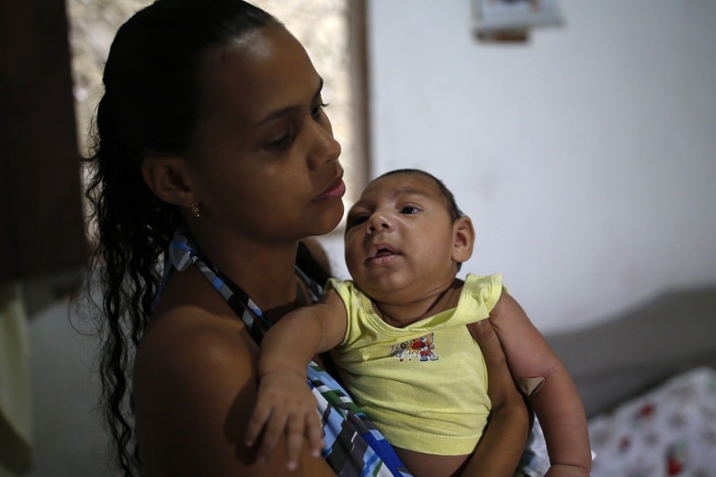
A team of researchers suspect they have discovered how the Zika virus probably causes microcephaly in fetuses. The virus selectively infects cells that form the brain’s cortex, or outer layer, making them more likely to die and less likely to divide normally and make new brain cells.
They say their experiments also suggest the highly susceptible lab-grown human stem cells they used could be employed to screen for drugs that protect the cells or ease existing infections.
READ MORE: Hong Kong faces greater threat from Zika virus with arrival of summer
It’s very telling that the cells that form the cortex are potentially susceptible to the virus, and their growth could be disrupted by the virus
“Studies of fetuses and babies with the telltale small brains and heads of microcephaly in Zika-affected areas have found abnormalities in the cortex, and Zika virus has been found in the fetal tissue,” said Dr Guo-li Ming, a professor of neurology, neuroscience, and psychiatry and behavioural science at the Institute for Cell Engineering at Johns Hopkins University in the United States. “While this study doesn’t definitely prove that Zika virus causes microcephaly, it’s very telling that the cells that form the cortex are potentially susceptible to the virus, and their growth could be disrupted by the virus.”
READ MORE: The Zika virus explained: why the world is not taking chances with birth defect-linked disease spread by mosquitoes
In 2015, the Zika virus began spreading throughout the Americas and a potential link was seen between the virus and a significant increase in cases of fetal microcephaly, as well as other neurological abnormalities. This connection and the proliferation in cases led to the World Health Organisation declaring Zika virus an international public health emergency.

Results of the experiments, conducted by researchers at Johns Hopkins, Florida State University, and Emory University, also in the US, are described online in the journal Cell Stem Cell.
READ MORE: Wiping out the mosquito species carrying the Zika virus is a good case for gene modification
In a quickly executed study that reflects the global public health threat posed by Zika, the researchers compared its effect on cells known as cortical neural progenitor cells to two other cell types: induced pluripotent stem cells and immature neurons. Induced pluripotent stem cells are made by reprogramming mature cells, and can give rise to any cell type in the body, including cortical neural progenitor cells. Cortical neural progenitor cells in turn give rise to immature neurons.